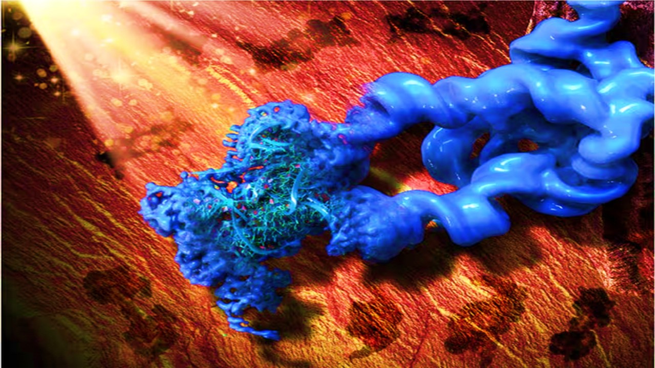
The realm of drug discovery and development has undergone a remarkable transformation in the past decades, primarily fueled by the integration of computational methods into the process. Among the pivotal areas benefiting from these advancements is the development of kinase inhibitors (KIs) as targeted anticancer drugs. Since the groundbreaking approval of imatinib in 2001, protein kinases have emerged as prime targets for innovative drug design in the fight against cancer. This article delves into the sophisticated world of in silico methods employed for designing kinase inhibitors, focusing on their application as potent anticancer agents.
In Silico Methods Unveiled
The surge in kinase-targeted drug development owes much of its success to computational techniques that facilitate the exploration of structural attributes pivotal for efficacious interaction between kinases and their ligands. This is where in silico methods come into play, with two primary approaches dominating the landscape of computer-aided drug design (CADD): structure-based drug design (SBDD) and ligand-based drug design (LBDD).
Structure-Based Drug Design (SBDD)
Structure-Based Drug Design (SBDD) is a powerful approach that capitalizes on the three-dimensional structural information of biological molecules to drive the rational design of potential drug candidates. In the context of kinase inhibitors, SBDD has proven instrumental in the development of targeted therapies against a wide array of cancers. SBDD leverages the intricate understanding of the structural characteristics of kinases to design compounds that interact optimally with their active sites, inhibiting their function and thereby impeding aberrant cellular signaling.
Molecular Docking: Probing Ligand-Binding Interactions
At the heart of SBDD lies molecular docking, a computational technique that provides invaluable insights into the binding process between a ligand (prospective drug) and a protein’s active site. In the case of kinase inhibitors, molecular docking simulations involve virtually positioning potential drug candidates within the three-dimensional structure of the kinase’s active site. By analyzing various conformations and orientations, the simulation predicts how well a ligand fits into the active site and identifies the specific interactions it forms with the kinase’s residues.
This process helps to identify ligands that possess the potential to form stable interactions with the kinase’s active site residues, increasing the likelihood of inhibiting the kinase’s activity. Docking also offers estimations of binding affinities, enabling the ranking of ligands by their predicted potency.
Structure-Based Virtual Screening: Scanning Compound Libraries
Another crucial SBDD technique is structure-based virtual screening (SBVS), which involves the systematic screening of large compound libraries against the three-dimensional structure of a target kinase. Unlike traditional high-throughput screening, which involves physically testing each compound, SBVS leverages computational resources to evaluate potential ligands virtually. By focusing on compounds that fit well into the kinase’s active site and display favorable interactions, SBVS accelerates the process of identifying promising lead compounds for further investigation.
Molecular Dynamics Simulations: Unveiling Dynamic Behavior
Molecular dynamics (MD) simulations are a pivotal tool within SBDD that provides insights into the dynamic behavior of both kinases and ligands. Kinases are inherently dynamic molecules that can shift between different conformations, which is particularly relevant for understanding their function and interactions. MD simulations computationally model the movement and behavior of atoms and molecules over time. For kinase inhibitors, MD simulations provide a deeper understanding of how ligands interact with the kinase in a dynamic environment, elucidating factors that influence binding stability and the potential for allosteric effects.
Additionally, MD simulations can explore the stability of ligand-bound kinase complexes over extended time scales, revealing the durability of their interactions and offering insights into potential resistance mechanisms that cancer cells might develop against kinase inhibitors.
Ligand-Based Drug Design (LBDD)
Ligand-Based Drug Design (LBDD) is a strategic approach that comes into play when detailed structural information about the target protein is not available or limited. In such cases, LBDD shifts the focus from the target protein’s structure to the known activity of ligands that interact with it. By capitalizing on the relationship between the structural features of these ligands and their biological activity, LBDD guides the design of new compounds with the potential to exhibit similar desired effects.
Quantitative Structure-Activity Relationship (QSAR) Modeling: Bridging Structure and Activity
A cornerstone of LBDD is the Quantitative Structure-Activity Relationship (QSAR) modeling, a method that establishes a mathematical relationship between the structural properties of a molecule and its biological activity. In the context of kinase inhibitors, QSAR modeling quantifies the relationship between specific molecular descriptors—such as electronic, steric, and hydrophobic properties—and the observed inhibitory activity against the target kinase. This mathematical model can then be applied to predict the activity of new, untested compounds.
QSAR models are particularly valuable in filtering out compounds that are unlikely to exhibit the desired activity, enabling researchers to prioritize the synthesis and testing of compounds that show the greatest potential. The predictive nature of QSAR modeling significantly accelerates the drug discovery process by directing resources toward the most promising candidates.
Advanced Forms of QSAR: Enhancing Predictive Power
3D-QSAR modeling represents a significant advancement in the field of LBDD. Traditional QSAR models utilize two-dimensional molecular descriptors to predict activity, but they often fail to capture the complexities of molecular interactions in three-dimensional space. 3D-QSAR models, such as Comparative Molecular Field Analysis (CoMFA) and Comparative Molecular Similarity Index Analysis (CoMSIA), address this limitation by incorporating three-dimensional information.
CoMFA and CoMSIA models map the three-dimensional spatial properties of ligands into mathematical equations, allowing for the consideration of both steric and electrostatic interactions. These models correlate these molecular properties to the observed biological activity, resulting in more accurate predictions. CoMFA, for example, creates fields that represent electrostatic and steric interactions at different points in the molecule, allowing researchers to understand how these properties affect the binding affinity.
Moreover, advanced 3D-QSAR models integrate quantum chemical parameters, enhancing the accuracy of predictions. These parameters consider the electronic structure and energy calculations of the molecules, providing a more comprehensive understanding of the interactions at play.
Harnessing Ligand-Based Methods for Kinase Inhibitor Design
In the relentless pursuit of effective anticancer therapies, Quantitative Structure-Activity Relationship (QSAR) models have emerged as crucial tools, enabling researchers to foresee how specific chemical attributes impact biological activity. These models serve as bridges between molecular structure and function, offering insights that are instrumental in the design and optimization of kinase inhibitors with potent anticancer potential.
Evolution of QSAR Models: From 2D to 3D Insights
The evolution of QSAR models mirrors the progress in our understanding of molecular interactions. At its inception, classical 2D-QSAR linked basic physicochemical parameters of compounds to their activity. This approach provided initial insights into how molecular properties influence biological response, but it lacked the ability to capture the nuances of three-dimensional interactions within protein binding sites.
A paradigm shift occurred with the introduction of more advanced 3D-QSAR models like CoMFA and CoMSIA. These models go beyond 2D descriptors by incorporating the spatial arrangement of atoms in molecules. CoMFA and CoMSIA harness complex algorithms to map steric and electrostatic properties across the ligand’s structure, creating comprehensive representations of how molecules fit and interact within protein binding pockets.
Merging QSAR and Molecular Dynamics: Redesigning for Enhanced Affinity
The synergy between QSAR and molecular dynamics (MD) simulations has catalyzed innovative advancements in kinase inhibitor design. One compelling example involves the redesign of second-generation Src kinase inhibitors. Src kinases, implicated in cancer progression, often develop resistance to inhibitors. By integrating QSAR insights with fragment-based drug discovery, researchers aimed to enhance the binding affinity of these inhibitors. QSAR guided the identification of modifications that could overcome resistance, and MD simulations predicted the stability of the redesigned inhibitors within the kinase’s active site.
Unveiling Structural Preferences for Drug Design
QSAR studies have also played a pivotal role in the creation of kinase inhibitors targeting specific proteins like IKK-β. Through rigorous analysis of chemical attributes and their impact on inhibitory activity, researchers identified structural preferences that correlate with enhanced kinase inhibition. This informed the design of potential drug candidates with greater affinity and selectivity.
Empowering Ligand-Based Virtual Screening (LBVS)
When the three-dimensional structure of a target protein remains elusive, researchers turn to Ligand-Based Virtual Screening (LBVS) as a powerful approach for identifying potential drug candidates. LBVS harnesses the known activity of ligands that interact with the target protein to guide the search for structurally similar compounds with desired biological effects. This strategy represents a pragmatic solution to the challenge of designing inhibitors without detailed structural insights.
Leveraging Known Active Compounds for Discovery
LBVS capitalizes on a fundamental principle: compounds with similar structures often exhibit similar biochemical properties. Researchers begin by identifying known active compounds—those that have shown efficacy against the target’s activity—often through experimental assays or literature searches. These compounds serve as templates for the design of virtual screening experiments.
Identifying Structurally Similar Compounds
In the LBVS process, researchers sift through vast molecular libraries containing a multitude of compounds. Using the known active compounds as guides, LBVS searches for molecules that share structural features with the template ligands. The assumption is that compounds with structural resemblance are more likely to interact with the target protein in a similar manner, potentially yielding the desired biological activity.
Pharmacophore-Based Virtual Screening: Unveiling Key Features
Pharmacophore-based virtual screening is a pivotal technique within LBVS. A pharmacophore is a spatial arrangement of chemical features that are essential for a ligand to interact with its target protein and elicit a specific biological response. By deciphering the key structural characteristics required for effective binding and activity, pharmacophore-based screening enhances the accuracy of compound selection.
Exemplifying LBVS with Tumor Progression Locus-2 (Tpl2) Kinase
A striking illustration of LBVS’s potency is its application in identifying potential inhibitors of the tumor progression locus-2 (Tpl2) kinase, a protein implicated in cancer development. With limited structural information available for Tpl2, LBVS and pharmacophore-based virtual screening were employed to identify compounds that aligned with the essential features for inhibiting Tpl2 activity.
This technique involves creating a pharmacophore model based on the known active ligands of Tpl2. The model encapsulates critical interactions required for effective inhibition. Subsequently, the molecular library is screened against this pharmacophore, rapidly identifying compounds that possess the necessary structural attributes to interfere with Tpl2 function.
Reshaping Precision Oncology Landscape
In silico methods have revolutionized the landscape of kinase inhibitor design as anticancer drugs. The fusion of structural insights and ligand-based strategies has enabled the discovery, design, and optimization of novel kinase inhibitors. As computing power continues to advance, virtual screening approaches hold promise in replacing traditional high throughput screening, offering an accelerated and cost-effective path to the development of potent anticancer agents. This integration of computational prowess with biochemical innovation stands poised to further reshape the landscape of targeted cancer therapy.
Subscribe
to get our
LATEST NEWS
Related Posts

Drug Discovery Biology
Governing Multi-Component Therapeutics: Andrea Small-Howard’s Systems Framework at GB Sciences, Inc.
A systems-driven analysis of Dr. Andrea Small-Howard’s leadership at GB Sciences, Inc., detailing how multi-component cannabinoid therapeutics, governance architecture, and AI-enabled discovery are converging to redefine translational drug development.

Drug Discovery Biology
Derisking Nucleic Acid Therapeutics: Sean Sullivan on CMC-Integrated Strategy at Arcturus Therapeutics
Sean Sullivan of Arcturus Therapeutics outlines how CMC-integrated strategy is derisking mRNA, oligonucleotide, and plasmid DNA therapeutics.

Read More Articles


Medicinal Chemistry & Pharmacology
April 14, 2026
Igor Nasonkin and Phythera Therapeutics: Moving Oncology Beyond Single Targets into Engineered Polypharmacologic Systems
Igor Nasonkin’s systems-driven approach at Phythera Therapeutics reframes oncology drug development from single-target inhibition to AI-enabled polypharmacologic network modulation using nature-derived molecular architectures.

Artificial Intelligence and Data Analytics
April 10, 2026
Inside Johnson & Johnson’s External Innovation Engine: Devin Swanson on Translating Integrated Discovery into Strategic Value
Devin Swanson’s leadership at Johnson & Johnson Innovative Medicines redefines external innovation as a tightly governed, AI-enabled translational system integrating multi-modal drug discovery, biomarker strategy, and capital-efficient execution.

Immunology & Oncology
April 9, 2026
From DMPK to Distributed Execution: Mehran F. Moghaddam’s Systems Strategy at OROX BioSciences, Inc.
A systems-level examination of how Mehran F. Moghaddam operationalizes DMPK, externalized R&D, and lipid-mediated therapeutics into a predictive, high-velocity biotech development architecture.
