Mitochondrial function declines with aging and is connected to abnormalities in mitochondria. Due to the buildup of mutations and oxidative damage brought on by reactive oxygen species (ROS), the volume, integrity, and functionality of mitochondrial DNA decline with advancing age. Mitochondrial dysfunction in older people is marked by diminished oxidative capability, decreased oxidative phosphorylation, decreased ATP synthesis, significantly increased ROS formation, and poor antioxidant defense. Age-related changes in mitochondrial dynamics and suppression of mitophagy, an autophagic process that eliminates defective mitochondria, cause a reduction in mitochondrial biogenesis. Mitochondrial function is further weakened and impaired by age-related anomalies in mitochondrial quality checks. The fraction of apoptotic cells rises in tissues that are older due to greater mitochondria-mediated programmed cell death.
Senescent Modifications in mtDNA
According to the mitochondrial theory of aging, mitochondrial DNA (mtDNA) ages more quickly than nuclear DNA with fewer effective repair mechanisms. Compared to nuclear DNA, mtDNA mutation rates can be up to 15 times higher. In fact, the buildup of mutations in mtDNA might very well cross a critical threshold and have negative effects, particularly in mitochondria where it is necessary to substitute components of the respiratory chain that are malfunctioning or damaged. Mitochondrial malfunction and increased ROS production can result from mutations in mtDNA that change the expression of oxidative phosphorylation (OxPhos) complexes. The mtDNA mutator mouse, a creature with mutant mtDNA polymerase, was created to demonstrate the significant risk of mtDNA mutations in aging. These mice developed accelerated aging symptoms due to a malfunctioning process in their mtDNA proofreading during replication, which led to the creation of several new mutations. The “vicious cycle” notion states that mtDNA mutations accumulate exponentially and should be linked to a noticeable increase in ROS generation. A linear trend in the acquisition of mtDNA mutations over the lifespan, however, has been observed in studies using mtDNA mutator mice. In the mtDNA mutator mice compared to the healthy animals, there were no appreciable differences in ROS generation or antioxidant enzyme activity. The “vicious cycle” explanation, which contends that mtDNA mutations and impaired OxPhos but not ROS generation are principally to blame for premature aging in the mtDNA mutator mice, is in fact substantially undermined by these data.
Moreover, mtDNA is extremely vulnerable to oxidative damage because of its vicinity to the ROS-producing elements of the respiratory chain and the lack of histones. In rat liver cells, mtDNA contained 16 times more 8-hydroxydeoxyguanosine than nuclear DNA, which is a sign of DNA oxidative stress. Rat liver and skeletal muscles showed significant age-related declines in the amount of mtDNA copies. These results imply that mtDNA quantity and structure may decrease with aging, resulting in abnormal production of electron transport chain proteins and a deterioration of the OxPhos machinery.
A Highly Active Powerplant Spells Stable ROS Supply
Denham Harmon first forth the free radical theory of aging in 1956. It has been proposed that a single factor, their shared propensity for producing ROS, may be used to explain the parallels between the toxic side effects brought on by exposure to ionizing radiation or hyperbaric oxygen. This idea fit well with Harmon’s own finding that respiration and, extrapolating from that, metabolic rate were inversely related to lifespan. All animals breathe, thus according to the free radical theory, aging is a reflection of the accumulated biomolecular harm brought on by the unavoidable and ongoing creation of ROS.
Electrons that occasionally leak or escape from the electron transport chain are the main natural source of ROS in cells. It is not difficult to imagine the creation of a negative feedback loop whereby a preliminary insult to the mitochondria can result in a higher incidence of ROS production that would, in turn, trigger additional harm. Serious harm to the components of this pathway would be anticipated to boost the rate of leakage. A buildup of damage to mitochondrial elements may therefore negatively impact the synthesis of ATP, maybe to the point where it compromises the health and functionality of cells.
The Unfortunate Lack of Endogenous DNA Repair Mechanisms
In this suggested redox damage cycle, the endogenous genome of the mitochondrion also participates. The genome of the ancient bacterium that served as the model for the present organelle can only be found in very small quantities in the mitochondrial genome. It is thought that the metabolite transfer between early eukaryotes and nearby bacteria evolved into a mutual dependence. The final result of this close metabolic symbiosis was endosymbiosis, which is when the smaller bacteria gets engulfed by its eukaryotic companion. The majority of the genes in the genome of the internalized bacteria were either deleted over time or were transferred to the nuclear DNA of the eukaryotic host, though not all of them. Currently, the human mitochondrion’s genome codes for 22 tRNAs, two ribosomal RNAs (one for each subunit), a number of the polypeptide components of complexes I, III, and IV of the electron transport chain, as well as fragments of the F1 and F0 ATPases.
The genes for the several types of monitoring and repair enzymes necessary to preserve the integrity of nuclear DNA are absent from the mitochondrial genome. As a result, when mutations take place, including those that negatively impact the electron transport chain, they are permanently incorporated into the genome of a mitochondrion. These changes and their effects not only last over time, but other mutations may also accumulate that further impair the functionality of the electron transport chain. One can also imagine a vicious loop where a mutation in the mitochondrial genome caused by ROS results in more ROS escape from the electron chain, which then causes additional genotoxicity and even more ROS permeability.
The mitochondrial hypothesis is still regarded as a potential contributor even though it is no longer thought to be a complete, all-encompassing explanation for all of the alterations connected to human aging and its comorbidities. The amount of the important electron carrier NAD(H) has been seen to decrease with aging. Furthermore, the central role that mitochondria play in programmed cell death (apoptosis) can be seen as providing evidence in favor of mitochondria as the cause of illness and senescence.
Powering Up Programmed Cell Death
Higher creatures are equipped with the ability to selectively remove cells that have undergone developmental alterations, such as those that occur often during embryogenesis, or that have been irreparably harmed. The apoptotic cell death pathway is activated by receptor-mediated signals during the remodeling of developing tissues. ROS, viral dsRNA, DNA damage, or heat shock may function as triggers in the case of damaged cells. These signals cause the permeability transition pore complex, which is part of the mitochondrial outer membrane, to expand, allowing the tiny (12.5 kDa or so), soluble electron carrier protein known as cytochrome C to exit into to the cytoplasmic portion of the cell. A multiprotein complex called the apoptosome forms here around cytochrome c as its central component. The proenzyme variants of the family of cysteine proteases known as caspases are the target of a series of proteolytic activation processes that commence when the apoptosome is assembled. The terminal caspases 3 and 7 degrade structural proteins in the cytoplasm and chromatin proteins in the nucleus, causing the pathologic cell to die and ultimately be phagocytosed. Many scientists are trying to figure out how to use the fact that there is an inherent, receptor-mediated cell death pathway as a way to target cancer cells and other dangerous cells for elimination.
Beyond Vision Lies Destruction
The light waves precisely underneath the blue end of the visible spectrum are referred to as ultraviolet (UV) radiation. They are strongly absorbed by organic molecules with aromatic rings or numerous, conjugated double bonds, even though the human eye cannot see them. The side chains of phenylalanine, tyrosine, and tryptophan; polyunsaturated fatty acids; heme groups; and various cofactors and coenzymes, such as flavins, cyanocobalamin, etc. are among these. The breakdown of covalent bonds in proteins, DNA, and RNA as well as the synthesis of thymine dimers in DNA, cross-linking of proteins, and the production of free radicals can all result from the absorption of this short wavelength, high intensity light.
Despite the fact that UV radiation can not permeate past the top few layers of epidermal cells, the great efficiency with which it is absorbed can cause damage to the skin to accumulate quickly. UV radiation is very mutagenic because the nucleotide bases of DNA and RNA are exceptionally susceptible to absorbing it. As a result, exposure to strong sunlight over an extended period of time can result in the buildup of many DNA lesions that surpass a cell’s inherent repair ability, resulting in the formation of myelomas—some of which rapidly multiply if left untreated.
Glycation Equals Destructive Cross-Link Formation
Protein and nucleotide amine groups potentially create an adduct through a process known as glycation when they are exposed to a reducing sugar like glucose. The first phase of this procedure is the creation of a Schiff base between the amine groups on proteins and other macromolecules and the aldehyde or ketone group of the sugar. Amadori products, which include a conjugated carbon-carbon double bond that can interact with the amino group on a nearby macromolecule, are created over time as a result of a sequence of conformational changes that glycated proteins go through. Covalent cross-links involving two proteins or other biological macromolecules are created as a result. Further glycation of these identical macromolecules can multiply the number of cross-links to incorporate more macromolecules. These cross-linked aggregates are also referred to as AGEs, or advanced glycation end products.
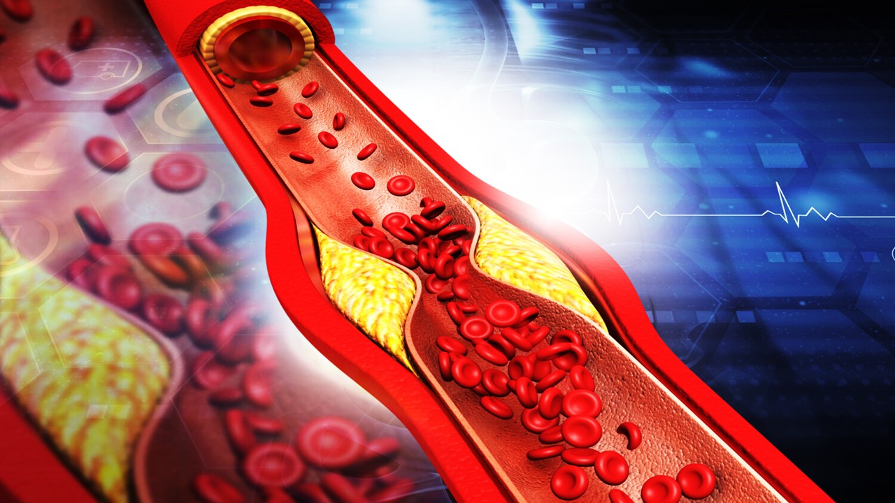
When long-lived proteins like collagen or β-crystallins are damaged, the effects of protein glycation on physiology can be particularly noticeable. Their continued presence increases the likelihood of numerous glycation and subsequent crosslinking events. The buildup of cross-links in the collagen network of vascular endothelial cells in blood vessels can result in a gradual elasticity reduction and hardening of the basement membrane, which in turn encourage the emergence of plaques. The final outcome is an increase in the heart’s burden. In the eye, the buildup of protein aggregates impairs the lens’ opacity and finally manifests as cataracts. Those with diabetes are more likely to develop AGEs because they have trouble controlling their blood sugar levels. In actuality, serum albumin and hemoglobin glycation serve as biomarkers for the detection of diabetes mellitus.
It Remains a Piece of the Aging Puzzle
Most eukaryotic cells get their energy mostly from their mitochondria, but they also get their free radicals primarily from them. The membranes, proteins, and DNA of a cell, as well as other components, can all be harmed by these reactive chemicals. As a result, they have long been thought to contribute to biological aging. Because mitochondria contain their own genetic material (mtDNA) and only a few number of DNA repair mechanisms available to them, they are one of the main targets for reactive oxygen species. The so-called mitochondrial theory of aging is based on the hypothesis that genetically damaged mitochondria build up over time and cause the aging phenotype by disrupting the energy budget.
This hypothesis has gained traction in the past couple of decades because of the identification of mitochondrial disorders and mtDNA deletions in aging species. In terms of the process of the accumulation of these deletions and their physiological significance, there are still a lot of unanswered concerns. Despite the fact that the mitochondrion’s part in the story of human aging and age-related pathologies seems solid, it is obvious that there are still many unwritten chapters in this tale.
Subscribe
to get our
LATEST NEWS
Related Posts

Chronic & Debilitating Diseases
Biochemical Topographies: Revealing Molecular Traces That Shape the Early Course of Diabetic Cardiomyopathy
Circulating biomarkers provide an essential roadmap for detecting the silent molecular progression of diabetic cardiomyopathy before it manifests as overt cardiac dysfunction.
Chronic & Debilitating Diseases
Silent Corpses, Dangerous Plaques: How Defective Cell Clearance Shapes Atherosclerosis Progression and Rupture
Failed clearance of apoptotic cells drives necrotic core expansion and inflammatory persistence, making defective efferocytosis a central determinant of atherosclerotic plaque vulnerability.


Read More Articles


Medicinal Chemistry & Pharmacology
April 14, 2026
Igor Nasonkin and Phythera Therapeutics: Moving Oncology Beyond Single Targets into Engineered Polypharmacologic Systems
Igor Nasonkin’s systems-driven approach at Phythera Therapeutics reframes oncology drug development from single-target inhibition to AI-enabled polypharmacologic network modulation using nature-derived molecular architectures.


Artificial Intelligence and Data Analytics
April 10, 2026
Inside Johnson & Johnson’s External Innovation Engine: Devin Swanson on Translating Integrated Discovery into Strategic Value
Devin Swanson’s leadership at Johnson & Johnson Innovative Medicines redefines external innovation as a tightly governed, AI-enabled translational system integrating multi-modal drug discovery, biomarker strategy, and capital-efficient execution.

Immunology & Oncology
April 9, 2026
From DMPK to Distributed Execution: Mehran F. Moghaddam’s Systems Strategy at OROX BioSciences, Inc.
A systems-level examination of how Mehran F. Moghaddam operationalizes DMPK, externalized R&D, and lipid-mediated therapeutics into a predictive, high-velocity biotech development architecture.