-Omics technologies have proven to be useful molecular tools across many therapeutic areas in facilitating qualitative and quantitative tissue analysis. Spatially Resolved Transcriptomics (SRT) is an example of -omics technology used in oncology to further investigate how the heterogeneity of the tumour microenvironment works. -Omics technologies like SRT methods continue to develop, with further integration into computational biology.
Introduction to -omics
The term -omics refers to a “field of study in biological sciences that ends with -omics, such as genomics, transcriptomics, proteomics, or metabolomics.” To clarify, genetics is a branch of biology which studies genetic function, variability and inheritance. A genome is the object of study within genetics; genomics is a more in depth analysis of genetics “which studies the structure, function, evolution, and mapping of genomes”.
In comparison with traditional biochemical methods, -omics technologies are more efficient and timesaving, used to produce large-scale data. -Omics technologies have important applications in biomedical and pharmaceutical research. These technologies have proven to be useful tools and “enable the exploration of the genome, transcriptome, and proteome more broadly with greater sensitivity and resolution”.
In the context of pharmaceutical research, -omics technologies can be used to provide solutions across drug development including target discovery and validation, drug toxicity and safety assessment.
High throughput -omics technologies can facilitate the analysis of vast libraries of molecules including genes and proteins in a very short period of time, which is useful for accelerating data-demanding analyses. In addition, the sensitivity, specificity and accuracy of the technology has enabled the identification of key biomarkers, a key part of molecular diagnostics which has seen -omics technology shift into precision medicine.
Next-generation sequencing (NGS) is an example of a large-scale DNA sequencing technology which characterises the entire genome for an organism. Whole genome sequencing is an important part of research for genetic-based diseases, allowing the compilation of germline variants increasing the risk for inherited diseases. NGS is a more cost-effective and time-efficient qualitative analysis in comparison to previous techniques, i.e. Sanger sequencing.
-Omics technology
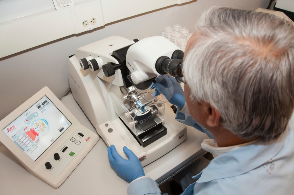
SRT is defined as a ”diverse set of technologies that encode gene expression measurements with information about where in the sample each observation occurred”. Microdissection-based methods are a complex group of SRT methods in which small sections of tissue or cells of interest are prepped, dissected and studied for cellular morphology and gene expression. Laser capture microdissection involves the staining of a small tissue section of a prepared glass slide to be imaged for morphology, and regions of interest include single cells using a microscope-guided laser.
In situ barcoding-based methods are another group of SRT techniques which enable spatial RNA quantification. One particular technique, Spatial Transcriptomics (ST) initially involves similar prep in which tissue is fixed, stained and then imaged. However the sequence of RNA quantification is inherently more complex involving permeabilisation, reverse transcription and cDNA library formation. The RNA sequence is mapped to specific tissue locations by aligning the histological image acquired at the beginning of the workflow to known spatial barcode locations.
Accurate qualitative analysis of cell morphology has proven the ability of SRT to interrogate tissue at an whole organism ‘-omics scale’. As a result, SRT is becoming an increasingly popular tool within oncology research.
SRT: Precision oncology
As described previously, SRT methods are an important part of qualitatively analysing the spatial variability of genetic expression within a sample. In cancer, there has been substantial evidence supporting spatial variations documented in pathological observations. Hence, SRT has become an important tool in oncology to study the genotypic variation under different tumour microenvironments (TME).
According to a journal publication, “the spatial association between genetically different cancer cells and blood vessels may be attributed to environmental adaptation, or the ability of cancer cells to modify their environments”. Therefore it is important to understand the spatial variability of the TME in identifying the factors driving tumour heterogeneity. Tumour heterogeneity refers to the variability of tumour cell morphology and phenotypic profiles in addition to differences in metabolism, expression and proliferation.
Previously, microdissection-based methods have been used for decades to study the clonal composition and cell-types of the TME with spatial resolution. Unfortunately, the process is highly labour-intensive and the complexity of dissection limits the throughput of these methods. Solid phase capture-based methods have since become an alternative choice with higher throughput and are suited for translational applications. Solid phase capture-based methods are primarily used to profile composition and spatial heterogeneity of the tumour microenvironment across tumour types.
In 2020, a publication investigated the spatial analysis of ligand-receptor interactions in skin cancer at genome-wide and single-cell resolution. The team used four complementary SRT technologies to measure gene expression in microarray-based ST-seq techniques (these study cellular interactions and disease mechanisms). A microarray is a tool used to measure the expression of pre-defined genes known as probes. In oncology, this technique has already been applied to prostate cancer and melanoma.
The results demonstrated that the expression of specific molecules in skin cancer was supported by RNAscope detection assays. In addition, the absolute quantification of the target’s genes through a separate PCR analysis, supported the choice of using an RNAscope assay for quantification.
Challenges
Despite their obvious potential for cancer studies, there remain a number of limitations using SRT methods. In the 2020 study, the team highlights how ST-seq still lacks single-cell resolution as well as the read quality that can be captured in each spot which is dependent on the tissue context.
In order to overcome the limited resolution of ST-sequencing, RNA-ISH is considered a more targeted approach to visualise interactions at single cell level within a region of interest. RNA-ISH, also known as RNA in situ hybridisation, is a “powerful tool to visualize target messenger RNA transcripts in cultured cells, tissue sections or whole-mount preparations”. This technique uses a novel probe design strategy and an amplification system to simultaneously amplify signals and suppress background. Furthermore, in the aforementioned study it is emphasised that there continue to be an absence of a comprehensive drug discovery pipeline studying ligand-receptor interaction from a cancer tissue section.
SRT methods are also moving towards computational techniques. For example, histological and SRT data has been used to train machine learning algorithms to predict histopathological annotations-based on gene expression data. The computational development of SRT has been observed across life science and the pharmaceutical industry, as noted in a recent Nature article in which a speaker foresees a phase of computational development around finding spatially variable genes or for finding trajectories that leverage the spatial information.
Charlotte Di Salvo, Lead Medical Writer
PharmaFeatures
Subscribe
to get our
LATEST NEWS
Related Posts
Bioinformatics & Multiomics
Mapping the Invisible Arrows: Unraveling Disease Causality Through Network Biology
What began as a methodological proposition—constructing causality through three structured networks—has evolved into a vision for the future of systems medicine.

Bioinformatics & Multiomics
Open-Source Bioinformatics: High-Resolution Analysis of Combinatorial Selection Dynamics
Combinatorial selection technologies are pivotal in molecular biology, facilitating biomolecule discovery through iterative enrichment and depletion.
Read More Articles
Myosin’s Molecular Toggle: How Dimerization of the Globular Tail Domain Controls the Motor Function of Myo5a
Myo5a exists in either an inhibited, triangulated rest or an extended, motile activation, each conformation dictated by the interplay between the GTD and its surroundings.


