Renal transplantation stands as the most frequently performed solid organ transplantation worldwide, a procedure critical to the survival and quality of life for patients suffering from end-stage kidney disease (ESKD). This surgical intervention has provided a viable alternative for thousands of patients annually, offering a path away from the chronic challenges of dialysis treatments, which, while life-sustaining, often come with lower quality of life and higher morbidity. Renal transplants, whether from living or deceased donors, afford these patients the best opportunity for a longer and healthier life, underscoring the value of this procedure within clinical medicine.
Despite its life-saving potential, renal transplantation is not without its own set of formidable challenges. Various postoperative complications can compromise patient outcomes, often leading to complex clinical scenarios that require nuanced management. Complications range from urological issues like urine leaks and obstructions to vascular challenges such as renal artery stenosis or thrombosis. Additionally, complications can arise from peritransplant fluid collections, including hematomas, lymphoceles, and abscesses, as well as risks related to herniation, calculous disease, and even neoplasms. Modern advancements in medical imaging, surgical practices, and postoperative care have mitigated some of these risks, significantly improving patient outcomes. However, the need for continued innovation to manage these complexities persists.
Technological Transformation: 3D Printing and Its Entrance into Transplant Surgery
The advent of 3D printing has created a paradigm shift in surgical practices, particularly within the high-stakes field of organ transplantation. Initially utilized outside the realm of healthcare for prototyping in engineering and manufacturing, 3D printing technology has since found a vital application in medicine, with particular significance in complex surgical procedures. In renal transplantation, 3D printing has introduced a new era of possibilities, allowing surgeons to better understand the intricate vascular structures they operate on, craft custom surgical tools, and prepare meticulously for complex anatomical challenges.
Three-dimensional printing, at its core, translates digital images into solid, tangible objects, achieved by sequentially layering materials until a complete, three-dimensional replica emerges. In medicine, this technology supports surgeons and clinicians by allowing them to visualize complex anatomical structures in ways that were previously unimaginable. The ability to produce physical models of organs or vasculature aids in education and preoperative planning, equipping surgeons with a more profound understanding of patient-specific anatomical intricacies. In the context of renal transplantation, where individualized approaches are often necessary, 3D printing allows for the precise customization that can enhance surgical precision and reduce intraoperative risks.
Beyond Supply Limitations: The Promise of Bioprinting in Overcoming Organ Shortages
The impact of 3D printing goes beyond mere surgical assistance; it also speaks to a potential solution to a critical bottleneck in renal transplantation: the shortage of available organs. Currently, only a small fraction of patients on transplant waiting lists receive a kidney within five years of being listed. Many patients experience health deterioration or premature death before a suitable organ becomes available. This shortage exacerbates the healthcare burden for ESKD patients, for whom dialysis represents an imperfect and often exhaustive alternative to transplantation. Bioprinting, an evolution of 3D printing technology, emerges here as a beacon of hope in addressing this disparity.
Bioprinting combines tissue engineering with 3D printing principles to create “bio-ink” structures that mimic natural tissue architecture. By utilizing biocompatible printers capable of constructing complex cellular matrices, bioprinting has introduced the potential for bioengineered tissues that could one day replace the reliance on organ donors. Although the technology has not yet reached the stage of producing fully functional kidneys, its progress is remarkable. Current applications allow for the creation of tissue-like structures with internal and external architecture that closely resemble natural kidney tissue, a process called biomimicry. This biomimetic approach supports cellular viability and function, creating promising grounds for further advancements in regenerative medicine.
A Revolutionary Prospect: Customizable Renal Transplants Through Bioprinting
Bioengineered kidney replacements have the potential to revolutionize renal transplantation by providing personalized, bio-printed organs tailored to individual patients’ needs. These advancements could bridge the gap between the demand and supply for kidneys, alleviating the strain on the healthcare system and offering new hope to patients who would otherwise face years on dialysis or indefinite waitlists. Individualized transplants created through bioprinting not only hold the promise of functional tissue replacement but also bring the possibility of circumventing immune rejection—a persistent issue in traditional transplantation that often necessitates lifelong immunosuppressive therapy.
The pathway to bioengineered kidneys, however, is intricate and laden with challenges. Research currently focuses on constructing viable, complex structures capable of sustaining cellular health and mimicking the functional characteristics of a human kidney. Efforts in bioprinting range from constructing glomeruli and other nephron structures to forming vascular systems capable of integrating with host vasculature post-transplant. Although complete kidney functionality is not yet a clinical reality, each step forward in bioprinting represents a leap toward achieving a sustainable, donor-independent solution to organ failure.
Patient Education: Empowering Through Visualization
In the world of renal transplantation, few things are as crucial as patient understanding. Complex surgical plans, unfamiliar medical terminology, and the inherent anxiety of major surgery can make it challenging for patients to fully grasp what lies ahead. This is where three-dimensional (3D) printing steps in as a transformative tool, bringing a new level of clarity to the patient experience.
Visualizing Complex Concepts
3D-printed models offer patients a tangible representation of their anatomy, allowing them to visualize not only their kidneys but also the surrounding structures that play a role in the surgical procedure. Such visual aids can demystify intricate details, helping patients understand their medical condition and the steps involved in their treatment. By transforming abstract concepts into concrete visuals, these models facilitate a deeper comprehension of what patients can expect before, during, and after the surgery.
Fostering Communication and Trust
The integration of 3D models into patient education promotes enhanced communication between healthcare providers and patients. When surgeons utilize these models in consultations, it fosters a collaborative environment where patients feel more engaged in their care. They can ask questions, express concerns, and gain reassurance through visual representation, ultimately leading to a stronger therapeutic alliance. This increased trust not only aids in informed consent but also empowers patients to take an active role in their healthcare journey.
Individualized Insights for Living Donors
For living donors, the benefits of 3D printing are particularly pronounced. Patient-specific models can demystify the donation process, providing clear visual guidance on how their anatomy will be affected. These models serve as a crucial tool for education, making the complexities of the procedure more accessible. By helping potential donors visualize the surgical process, healthcare providers can facilitate discussions that address their concerns and questions, ensuring they feel confident in their decision.
Enhancing Satisfaction and Understanding
Studies indicate that patients who interact with 3D-printed models demonstrate significant improvements in their understanding of their condition and the associated surgical procedures. For instance, following a model presentation, patients reported enhanced comprehension of kidney physiology, tumor characteristics, and the intricacies of the planned surgery. Such positive outcomes underscore the value of incorporating these innovative tools into clinical practice.
Preoperative Planning: A New Dimension in Transplantation
The advent of advanced 3D printing techniques, bolstered by detailed anatomic imaging, has ushered in a transformative era for preoperative planning in surgery. While 3D models have long been utilized across various medical fields, their application in transplantation is particularly promising. Numerous studies have focused on the role of 3D models specifically for preoperative planning in renal transplantation, highlighting their growing significance.
Understanding Anatomy for Optimal Outcomes
During kidney transplantation, the recipient’s pelvic anatomy plays a critical role in surgical feasibility and overall outcomes. The compatibility of the kidney graft with the recipient’s pelvic dimensions is essential, especially in cases such as polycystic kidney disease, where native kidneys can be substantially enlarged. Three-dimensional printed models are proving invaluable in this context, allowing surgeons to familiarize themselves with the surgical landscape before the operation.
For instance, researchers have developed anatomical 3D models representing both the donor’s kidney graft and the recipient’s pelvic cavity. These models provide surgeons the opportunity to practice vascular anastomoses in a controlled environment, enhancing their understanding of the anatomical relationships involved and facilitating discussions among the surgical team. The preoperative simulations conducted with these models closely mirror actual surgical procedures, significantly improving intraoperative navigation.
Tailoring Solutions for Pediatric Patients
The complexities of pediatric living donor kidney transplantation often hinge on anatomical considerations, such as size discrepancies between the graft and the recipient’s abdomen. In one notable case, a 3D-printed model helped avoid a mismatch between an incompatible kidney graft and a two-year-old recipient. This model captured critical abdominal structures, allowing the surgical team to devise a comprehensive preoperative plan and successfully execute a two-stage operation. Additionally, this technology enabled the identification of anatomical variations, particularly in cases with atypical vasculature, thus enhancing decision-making regarding surgical approaches and minimizing risks.
Enhancing Surgical Precision with Arterial Modeling
Arterial calcifications are a common concern among patients with end-stage kidney disease (ESKD) and can significantly impact the success of vascular anastomoses during transplantation. Some researchers explored this challenge by fabricating 3D models of the aortoiliac axis for recipients with varying degrees of arterial calcification. These models allowed surgeons to assess arterial feasibility preoperatively, even simulating the clamping of arteries to better understand the optimal sites for anastomosis. The qualitative assessment of these models revealed high scores for anatomical accuracy and usefulness in preoperative planning, underscoring the potential of 3D printing in enhancing surgical outcomes.
Streamlining Donor Surgery
The safety and rapid recovery of living donors remain paramount in transplantation, prompting innovations such as robotic-assisted nephrectomy as part of enhanced recovery protocols. The anatomical complexity, including the presence of multiple renal arteries or veins, directly influences the surgical approach. A recent study compared traditional surgical methods with 3D printing-assisted planning in living donor-transplant recipient pairs. The findings were striking: the use of 3D-printed models resulted in a higher diagnostic rate of vascular variations, reduced operation time, minimized intraoperative bleeding, and expedited recovery. These models facilitated tailored surgical strategies that optimized outcomes while reducing complications.
Supporting Complex Surgical Decisions
Three-dimensional printed kidney models have proven to be invaluable in planning partial nephrectomies, especially for complex renal tumors. These models not only enhance surgeons’ understanding of intricate anatomic details but also allow for tactile examination prior to surgery. At an international urological meeting, the utility of 3D models was positively reviewed, emphasizing their role in surgical planning and improved outcomes.
Bioprinting in Renal Regenerative Medicine: A Pathway to Innovation
Renal transplantation, while recognized as the gold standard for treating end-stage kidney disease (ESKD), represents a transitional solution rather than a definitive cure. This approach alleviates the burden of chronic dialysis but introduces its own challenges, such as the long-term effects of immunosuppression therapy. These therapies expose patients to increased risks of opportunistic infections and malignancies, complicating their health management. Alarmingly, up to 50% of kidney transplant recipients may lose their grafts within ten years due to chronic allograft nephropathy. This underscores the need for innovative strategies to address the underlying issues of kidney disease.
The intricacy of kidney architecture, comprised of over 20 distinct cell types, presents formidable challenges for effective bioprinting. However, the potential of 3D bioprinting in achieving precise anatomical structures offers a promising avenue for mimicking kidney function. Two principal strategies have emerged in organ bioprinting: scaffold-based and scaffold-free approaches. Although 3D bioprinting is in its infancy, particularly concerning the development of complex renal structures, significant advancements are underway.
Innovations in 3D Bioprinting for Renal Applications
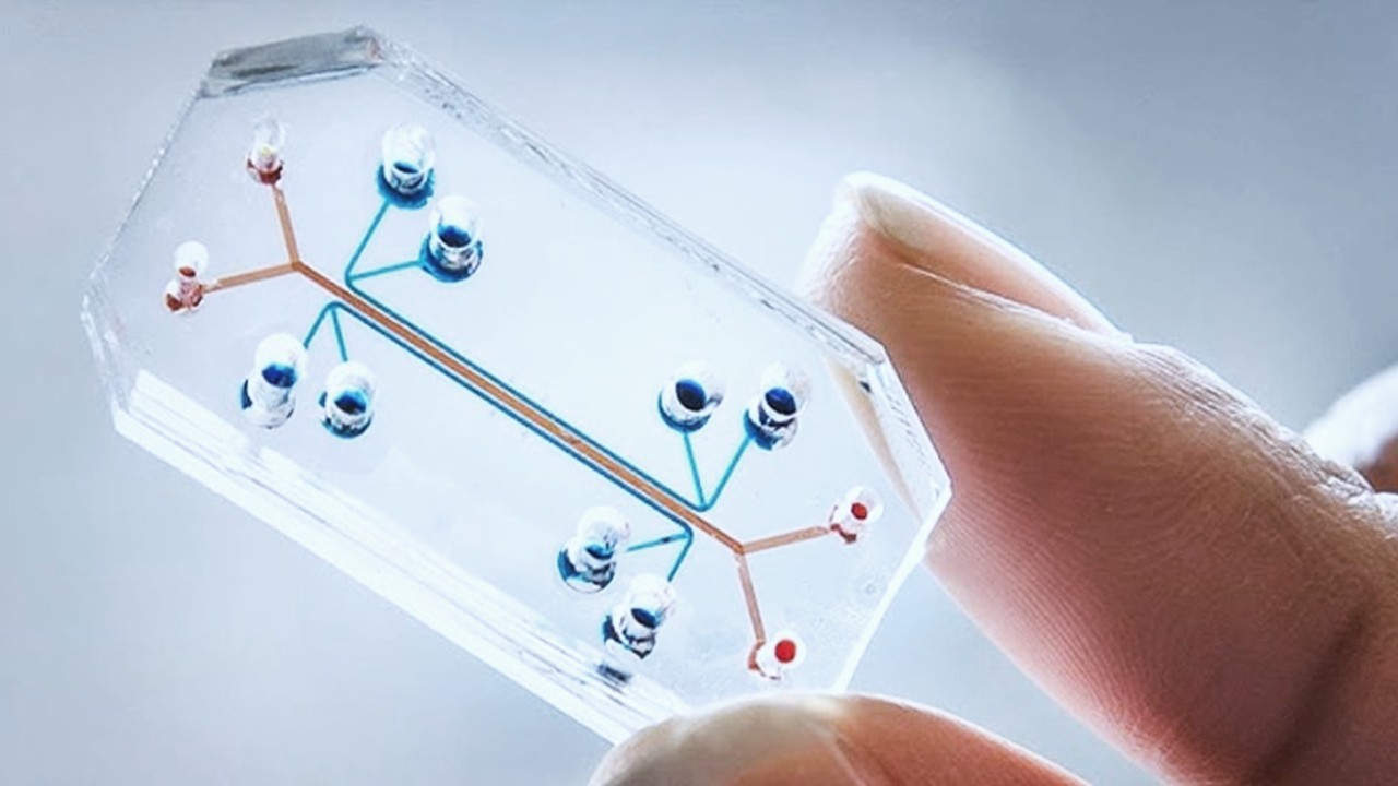
Recent studies have illuminated the potential of 3D bioprinting in the development of renal tissues and cultures. The first groundbreaking study in 2016 successfully bioprinted convoluted renal proximal tubules in vitro. This research highlighted the ability to create a perfusable microfluidic chip housing renal proximal tubules, which were fully embedded in an extracellular matrix. The architecture allowed for cell viability and functionality to be sustained for over two months, marking a significant advancement in renal modeling.
Building on this foundation, subsequent studies have focused on vascularized models that demonstrate active solute reabsorption and the potential for disease modeling. Proximal tubules, as primary sites of nephrotoxicity, are pivotal in drug screening processes. Research has demonstrated the effectiveness of 3D bioprinted renal constructs in simulating drug-induced tubular damage and assessing nephrotoxicity, providing a critical tool for early-stage drug development.
The Role of Bio-Inks in Bioprinting Success
The choice of bio-ink is crucial in ensuring the long-term viability and functionality of bioprinted renal constructs. For instance, a study highlighted the importance of incorporating various cell types and growth factors into bio-inks to promote healthy cellular interactions and maintain metabolic activity. Such formulations have shown promising results in supporting the growth and maturation of renal cells, with significant implications for regenerative medicine.
Moreover, the capacity to fabricate renal organoids using human pluripotent stem cells represents a breakthrough in creating more effective models for studying renal disease. These organoids have proven superior to traditional 2D cultures in predicting drug-induced nephrotoxicity, showcasing the power of 3D bioprinting in advancing renal research.
Bioprinting in Regenerative Strategies for ESKD
Beyond creating transplantable kidneys, 3D bioprinting has the potential to significantly enhance regenerative medicine for ESKD. Even a modest restoration of renal function—around 10%—could liberate patients from the clutches of dialysis, vastly improving their quality of life. Recent research has explored innovative approaches, such as developing autologous omentum patches to treat ESKD, demonstrating promising outcomes in reducing renal damage and promoting recovery.
In another groundbreaking study, researchers utilized decellularized porcine kidneys to produce hybrid bio-inks for 3D bioprinting, effectively creating vascularized tubular renal tissues. Transplanting these grafts into chronic renal disease models yielded promising results, including decreased markers of renal damage and enhanced neovascularization.
A Bright Future for Renal Bioprinting
The landscape of renal regenerative medicine is rapidly evolving, with 3D bioprinting poised to redefine treatment paradigms for ESKD and enhance transplant outcomes. By addressing the multifaceted challenges of kidney disease through innovative bioprinting techniques, researchers are paving the way for more effective therapies and ultimately transforming patient care. As the field continues to advance, the prospect of creating fully functional renal structures may soon transition from the realm of possibility to clinical reality, offering hope for millions affected by renal disorders.
Study DOI: https://doi.org/10.3390/jcm12206520
Engr. Dex Marco Tiu Guibelondo, B.Sc. Pharm, R.Ph., B.Sc. CpE
Editor-in-Chief, PharmaFEATURES
Register your interest [here] at Proventa International’s Clinical Operations and Clinical Trials Supply Chain Strategy Meeting this 14th of November 2024 at Le Meridien Boston Cambridge, Massachusetts, USA to engage with thought leaders and like-minded peers on the latest developments in the clinical space and regulatory affairs.

Subscribe
to get our
LATEST NEWS
Related Posts

AI, Data & Technology
Precision in Three Dimensions: A Novel Approach to Tumor Resection and Reconstruction of the Femoral Trochanter
The integration of digital modeling and personalized guides into the surgical workflow transforms the execution of tumor resection and reconstruction.

AI, Data & Technology
Blueprint for the Future: Establishing Rigorous Standards for Medical AI Data
Medical AI requires not just vast datasets but datasets of impeccable quality.
Read More Articles
Myosin’s Molecular Toggle: How Dimerization of the Globular Tail Domain Controls the Motor Function of Myo5a
Myo5a exists in either an inhibited, triangulated rest or an extended, motile activation, each conformation dictated by the interplay between the GTD and its surroundings.


