From Flat Cells to 3D Precision: The Rise of Lung Organoids
The evolution of lung organoids marks a transformative era in respiratory disease modeling. These miniature, three-dimensional structures replicate the complexity of human lung tissue, offering unparalleled insights into pulmonary biology. Traditional two-dimensional (2D) cell cultures and animal models, while invaluable, fall short in mimicking the cellular intricacies of human lungs. Enter lung organoids, derived from stem cells and engineered to mimic the structural and functional attributes of the respiratory system.
Lung organoids bridge the gap between 2D cell culture and living organisms. Unlike 2D cultures, which lack cellular diversity and spatial organization, lung organoids recreate the multilayered complexity of lung tissue. Their development relies on adult stem cells (ASCs) or pluripotent stem cells (PSCs), both of which possess the remarkable ability to self-organize into lung-like structures. These organoids have demonstrated potential in modeling respiratory diseases such as COVID-19, respiratory syncytial virus (RSV), and pulmonary fibrosis, establishing themselves as indispensable tools in translational research.
Despite these advances, lung organoids face significant challenges. Their immaturity, cellular heterogeneity, and limited functional capability compared to native lung tissue necessitate continued innovation. Researchers are addressing these limitations by co-culturing organoids with endothelial cells, mesenchymal cells, and immune cells, as well as integrating bioengineering platforms like air-liquid interfaces (ALI), microfluidic chips, and hydrogels. These advancements are paving the way for a more accurate understanding of respiratory diseases and their underlying mechanisms.
Building Complexity: Cellular Niches and Co-Culture Systems
One of the most compelling aspects of lung organoid research is the integration of cellular niches, which enhances organoid complexity and functionality. Endothelial cells, mesenchymal cells, and immune cells play pivotal roles in mimicking the native lung microenvironment, enabling researchers to create organoids that better reflect human physiology.
Endothelial cells, essential for vascularization, are critical in forming functional alveolar-capillary barriers. Studies have demonstrated that co-culturing endothelial cells with lung epithelial cells results in perfusable vascular networks, enhancing gas exchange efficiency. Similarly, mesenchymal cells provide structural support and influence alveolar epithelial cell differentiation. By embedding lung organoids in extracellular matrix hydrogels enriched with mesenchymal cells, researchers have improved the maintenance of alveolar stem cell populations and their differentiation potential.
Immune cells, another vital component of the lung microenvironment, are integral to studying disease progression. Co-culturing alveolar macrophages with lung organoids has provided insights into macrophage-epithelium crosstalk and immune responses to respiratory pathogens. These multicellular co-culture systems have been particularly useful in modeling infections such as SARS-CoV-2 and Mycobacterium tuberculosis, demonstrating the importance of incorporating niche-specific cells in organoid research.
Engineering Precision: Bioengineering Platforms in Organoid Culture
The development of advanced bioengineering platforms has significantly enhanced the structural and functional fidelity of lung organoids. Air-liquid interfaces (ALI), microfluidic chips, and hydrogels have been instrumental in creating physiologically relevant culture environments.
The ALI system, where the apical side of the cells is exposed to air while the basal side remains submerged in a nutrient-rich medium, mimics the native lung’s spatial and functional characteristics. This method has been particularly effective in cultivating organoids with differentiated airway and alveolar epithelial cells. ALI-based systems have also been used to study viral infections, such as SARS-CoV-2, by replicating host-pathogen interactions in a controlled environment.
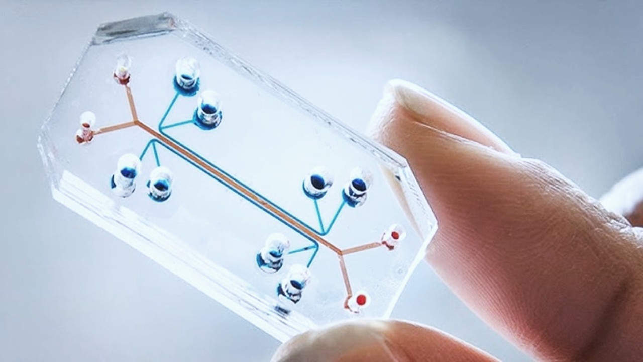
Microfluidic chips, often referred to as “lungs-on-a-chip,” enable researchers to simulate the mechanical forces and fluid dynamics of respiration. These devices integrate lung organoids into microengineered systems, facilitating studies on alveolar-capillary interactions, drug delivery, and disease progression. Additionally, hydrogels, both natural and synthetic, provide a three-dimensional scaffold that supports organoid growth. Decellularized lung extracellular matrix hydrogels, for instance, have been shown to enhance alveolar epithelial cell differentiation and proliferation, underscoring their potential in regenerative medicine.
Disease in a Dish: Modeling Respiratory Infections
Lung organoids have revolutionized the study of respiratory infections, offering a high-fidelity model to investigate pathogen-host interactions. SARS-CoV-2, the virus responsible for COVID-19, has been extensively studied using lung organoids. These models have revealed how the virus targets alveolar epithelial cells and disrupts lung function. Advanced infection platforms, such as apical-out organoid cultures and ALI systems, have further streamlined the study of viral entry, replication, and immune responses.
Respiratory syncytial virus (RSV) and influenza A virus have also been studied using lung organoids. These infections, which pose significant health risks to vulnerable populations, have been shown to cause cell-specific damage, such as ciliated cell disruption and alveolar epithelial cell apoptosis. Lung organoids have not only elucidated the mechanisms of these infections but also facilitated drug screening and therapeutic development.
Bacterial pathogens, including Mycobacterium tuberculosis and Streptococcus pneumoniae, have been investigated using lung organoid models. These studies have highlighted the complex interplay between bacterial pathogens and lung epithelial cells, providing valuable insights into host-pathogen dynamics and potential therapeutic targets.
Fibrotic Transformations: Modeling Chronic Lung Diseases
Chronic lung diseases, such as idiopathic pulmonary fibrosis (IPF) and chronic obstructive pulmonary disease (COPD), present unique challenges in disease modeling. Lung organoids offer a promising platform to study these conditions, capturing the cellular and molecular changes associated with fibrosis and inflammation.
IPF, characterized by the progressive scarring of lung tissue, has been modeled using organoids treated with transforming growth factor-beta (TGF-β). These models have replicated key features of the disease, including epithelial-mesenchymal transition and extracellular matrix deposition. Similarly, COPD organoids derived from patient cells have demonstrated disease-specific phenotypes, such as goblet cell hyperplasia and reduced ciliary function.
Advancements in genetic engineering, such as CRISPR/Cas9, have enabled the creation of organoids with specific genetic mutations linked to lung diseases. For instance, lung organoids with mutations in the CFTR gene have been used to study cystic fibrosis, paving the way for personalized medicine and gene therapy.
The Road Ahead: Challenges and Opportunities
While lung organoids have achieved remarkable progress, challenges remain. The immaturity of organoids, limited scalability, and the difficulty in replicating chronic disease conditions are barriers that need to be addressed. Integrating advanced technologies, such as 3D bioprinting and organoid-on-a-chip systems, holds promise for overcoming these limitations.
Moreover, the standardization of organoid culture protocols and the development of automated platforms will enhance reproducibility and scalability, enabling broader applications in drug discovery and regenerative medicine. By combining cellular engineering, bioengineering platforms, and advanced analytics, lung organoids are poised to revolutionize respiratory research and clinical practice.
In the quest to understand and treat pulmonary diseases, lung organoids stand at the forefront, offering a window into the complexities of the respiratory system. As this technology continues to evolve, it promises to deliver transformative insights into human lung biology, with far-reaching implications for global health.
Study DOI: https://doi.org/10.1177/20417314241232502
Engr. Dex Marco Tiu Guibelondo, B.Sc. Pharm, R.Ph., B.Sc. CpE
Editor-in-Chief, PharmaFEATURES

Subscribe
to get our
LATEST NEWS
Related Posts

Molecular Biology & Biotechnology
Myosin’s Molecular Toggle: How Dimerization of the Globular Tail Domain Controls the Motor Function of Myo5a
Myo5a exists in either an inhibited, triangulated rest or an extended, motile activation, each conformation dictated by the interplay between the GTD and its surroundings.

Drug Discovery Biology
Unlocking GPCR Mysteries: How Surface Plasmon Resonance Fragment Screening Revolutionizes Drug Discovery for Membrane Proteins
Surface plasmon resonance has emerged as a cornerstone of fragment-based drug discovery, particularly for GPCRs.
Read More Articles
Designing Better Sugar Stoppers: Engineering Selective α-Glucosidase Inhibitors via Fragment-Based Dynamic Chemistry
One of the most pressing challenges in anti-diabetic therapy is reducing the unpleasant and often debilitating gastrointestinal side effects that accompany α-amylase inhibition.


