Lipids as Molecular Sentinels: A New Frontier in Cancer Screening
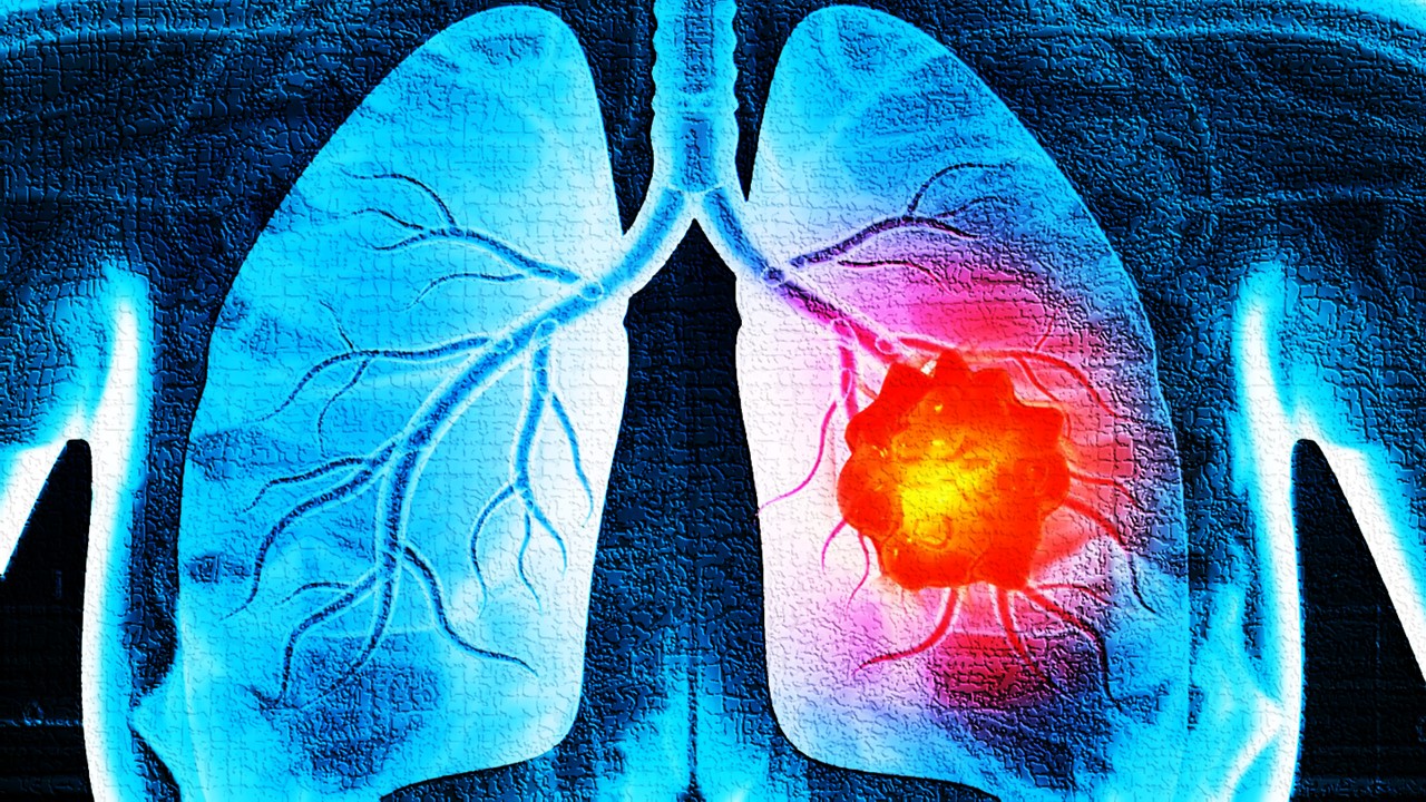
Cancer remains a formidable challenge in modern medicine, with early detection being a critical determinant of survival. Advances in diagnostic tools have expanded the frontier of detection techniques, but the non-invasive, cost-effective, and high-throughput analysis of peripheral blood emerges as the most patient-friendly and scalable option. Within this landscape, lipids, vital biomolecules involved in cellular structure, signaling, and energy storage, are gaining traction as biomarkers. Variations in their concentrations often reflect the biochemical underpinnings of disease.
A recent study leverages lipidomic profiling to identify cancer-specific plasma signatures. Researchers applied ultrahigh-performance supercritical fluid chromatography coupled with mass spectrometry (UHPSFC/MS) to dissect the plasma lipidome of patients with breast, kidney, and prostate cancers. Their findings reveal that lipid profiles not only differentiate cancer patients from healthy individuals but also hold promise for creating a universal biomarker panel. This development marks a significant stride toward redefining the standards for cancer screening, enabling earlier and more accurate diagnoses.
Through meticulous statistical modeling and validation, the study highlights seven key lipids—CE 16:0, Cer 42:1, LPC 18:2, PC 36:2, PC 36:3, SM 32:1, and SM 41:1—as potential biomarkers. These lipids capture the pathological nuances of cancer metabolism, pointing to a future where routine blood tests could replace invasive biopsies for initial cancer screening.
The Lipidomic Landscape: Charting Disease-Specific Signatures
Lipids, as integral cellular components, provide a snapshot of physiological states. The lipidomic profiles of plasma samples from over 480 individuals—comprising healthy controls and patients with breast, kidney, and prostate cancers—were analyzed in the study. The goal was to uncover lipid patterns that distinguish pathological states from health. Employing UHPSFC/MS as the analytical tool of choice, researchers quantified 138 lipids, encompassing glycerolipids, glycerophospholipids, and sphingolipids.
Using multivariate data analysis (MDA), the team visualized the separation between cancer and control samples. Principal component analysis (PCA) identified clusters, while orthogonal projection to latent structures discriminant analysis (OPLS-DA) confirmed the lipidomic distinctions between patient and control groups. Interestingly, specific lipid classes—such as sphingolipids and glycerophospholipids—emerged as dominant players in breast and prostate cancers, while nonpolar lipids like cholesteryl esters were more relevant for kidney cancer.
This systematic dissection revealed a recurrent pattern: plasma lipids were consistently downregulated in cancer patients. These reductions likely reflect the metabolic shifts occurring in cancer cells, which divert lipid resources for membrane synthesis, signaling, and energy production. The study’s robust analytical pipeline, coupled with its focus on method repeatability, ensures that these findings are not artifacts of measurement but reliable indicators of disease state.
Seven Lipids to Watch: The Biomarker Panel
The study zeroed in on seven lipid species that emerged as the most significant biomarkers across all cancer types. These lipids not only displayed statistically significant differences between patient and control groups but also maintained their diagnostic relevance across analytical methods and validation phases. Their biological roles shed light on why they may be altered in cancer:
CE 16:0 (Cholesteryl Ester 16:0). A reservoir of palmitic acid, crucial for lipid synthesis, signaling, and cancer cell proliferation. Cancer cells may deplete circulating CE 16:0 to meet their metabolic demands.
Cer 42:1 (Ceramide 42:1). A sphingolipid implicated in cell growth and apoptosis. Dysregulation of ceramides in cancer suppresses apoptotic pathways, enabling unchecked proliferation.
LPC 18:2 (Lysophosphatidylcholine 18:2). A proinflammatory lipid that modulates immune responses and promotes tumor growth. LPC depletion in plasma may reflect its diversion to the tumor microenvironment.
PC 36:2 and PC 36:3 (Phosphatidylcholine 36:2 and 36:3). Major components of cellular membranes, essential for cancer cell survival and metastasis. Their altered metabolism is a hallmark of cancer progression.
SM 32:1 and SM 41:1 (Sphingomyelin 32:1 and 41:1). Sphingolipids involved in signaling pathways. Cancer-induced dysregulation of sphingomyelins reflects the metabolic rewiring of tumor cells.
These lipids encapsulate the metabolic reprogramming characteristic of cancer, making them promising candidates for a unified biomarker panel. However, further studies are needed to validate their clinical utility across diverse populations and cancer subtypes.
Refining the Toolbox: Lipidomics and Analytical Rigor
The reliability of lipid-based diagnostics hinges on the reproducibility of results across different methodologies and timeframes. To this end, the study re-analyzed plasma extracts months apart, ensuring the stability and robustness of UHPSFC/MS measurements. Additionally, shotgun mass spectrometry, an independent analytical technique, was employed to verify the findings. Both approaches corroborated the downregulation of key lipids in cancer patients.
Statistical models were further stress-tested by reducing the number of lipids included in MDA. Remarkably, even with as few as seven lipid species, the models retained high specificity and accuracy. This simplification paves the way for streamlined clinical assays, reducing complexity without sacrificing diagnostic power.
The study also explored gender-specific differences in lipid profiles, given the inherent biological variations. Separate models for men and women enhanced predictive accuracy, particularly for kidney cancer. These insights underscore the importance of tailoring lipidomic analyses to account for demographic and physiological diversity.
Implications for Early Detection: From Bench to Bedside
The potential of lipidomics extends far beyond academic curiosity. By identifying plasma lipid profiles as reliable indicators of cancer, this study lays the groundwork for practical applications in population screening. Routine blood tests could incorporate lipidomic panels, enabling the early detection of cancers like breast, kidney, and prostate long before symptoms manifest.
The benefits of such an approach are manifold. First, lipidomics offers a minimally invasive alternative to current diagnostic methods, which often rely on imaging or biopsies. Second, its scalability makes it suitable for large-scale screening programs. Finally, by focusing on molecular changes rather than anatomical abnormalities, lipidomics promises higher sensitivity in detecting early-stage cancers.
However, challenges remain. The observed lipid alterations may not be exclusive to cancer, as other pathological conditions could influence plasma lipid levels. Future studies must address this specificity by comparing cancer patients with those suffering from non-malignant diseases. Additionally, the integration of lipidomics into clinical workflows will require standardized protocols, inter-laboratory reproducibility, and regulatory approval.
Toward a Lipid-Based Revolution in Oncology
This study marks a pivotal step in the quest for non-invasive, accurate, and early cancer diagnostics. By illuminating the unique lipidomic signatures of breast, kidney, and prostate cancers, it underscores the potential of plasma lipids as molecular sentinels. The identification of a seven-lipid biomarker panel offers a tantalizing glimpse of a future where routine blood tests could serve as the first line of defense against cancer.
As research progresses, the hope is that lipidomics will move from the realm of discovery to clinical application, transforming the way we detect and manage cancer. This lipid-based revolution has the potential to not only improve patient outcomes but also redefine the standard of care in oncology, one lipid at a time.
Study DOI: https://doi.org/10.1038/s41598-021-99586-1
Engr. Dex Marco Tiu Guibelondo, B.Sc. Pharm, R.Ph., B.Sc. CpE
Editor-in-Chief, PharmaFEATURES

Subscribe
to get our
LATEST NEWS
Related Posts

Immunology & Oncology
The Silent Guardian: How GAS1 Shapes the Landscape of Metastatic Melanoma
GAS1’s discovery represents a beacon of hope in the fight against metastatic disease.
Immunology & Oncology
Resistance Mechanisms Unveiled: The Role of Glutathione S-Transferase in Cancer Therapy Failures
Understanding this dual role of GSTs as both protectors and accomplices to malignancies is central to tackling drug resistance.
Read More Articles
Myosin’s Molecular Toggle: How Dimerization of the Globular Tail Domain Controls the Motor Function of Myo5a
Myo5a exists in either an inhibited, triangulated rest or an extended, motile activation, each conformation dictated by the interplay between the GTD and its surroundings.


