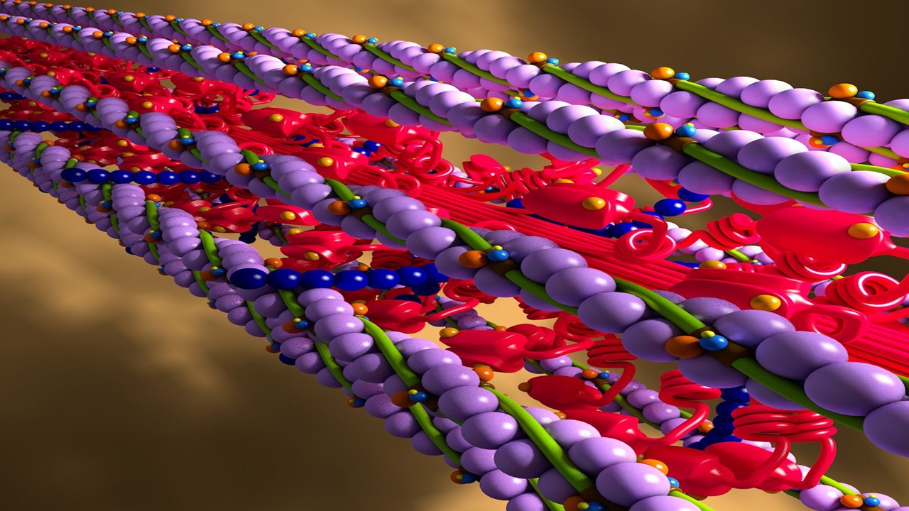
Creative Proteomics subsidiary Cytokine provides customized investigation and analysis solutions to federal agencies, academia, and scientists in the pharmaceutical and biotechnological sectors. The company’s Cancer Cytokines Assay, which can identify multiple cytokine levels in a single sample, was just released on the first week this month, according to the product manager for the business.
The Importance of Cytokines in Fighting Cancer
Several cytokines and growth factors are released by cancer cells during the tumor-forming process, tweaking adjacent cellular components to create a tumor microenvironment (TME). Endothelial cells, fibroblasts, and invading inflammatory cells are abundant in malignant cells. According to research, cytokines are crucial to the emergence and growth of malignancies.
To harness the immune system’s ability to combat cancer and remove aberrant cells, as well as to limit or stop their progression, myriad treatments have been created. In order to hasten the development of molecular pathogenesis and condition immunotherapy clinical works, Creative Proteomics delivers cancer-associated factor analysis.
Cytokines Involved in Cancer
Tumor cell-derived VEGF acts on endothelial cells to promote angiogenesis, tumor growth, invasion, and metastasis. At tumor sites, monocytes differentiate into tumor-associated macrophages capable of producing growth factors, proteases, angiogenic mediators, cytokines, and chemokines such as CCL2 (MCP-1), CCL5 (RANTES). Interleukin (IL-2), IL-6, IL-10, IL-12, interferon (IFN), tumor necrosis factor (TNF), transforming growth factor (TGF) play a crucial role in promoting tumor progression and anti-tumor. Chemokines such as CXCL10 (IP-10) and CXCL9 may inhibit the development of cancer. There are also many adhesion molecules expressed by different types of tumor cells that are often key factors for metastatic malignant tumors, such as ICAM-1, ICAM-3, VCAM-1, and selectins.
Cytokine Assay Technology
Cytokine uses the Luminex cytokine detection platform, which uses fluorescently encoded microspheres with specific antibodies to detect multiple target molecules simultaneously. The Luminex cytokine assay platform offers multiple detection, short experiment time, high sensitivity, saves samples, and time.
Principle of Luminex Multiplex Assays
Superparamagnetic 6.5-micron microspheres with a magnetic core and polystyrene surfaces are used in Luminex technology. A specific ratio of red and infrared fluorophores is used to dye the beads’ interiors, creating 100 different spectral signature microspheres. A 96-well microtiter plate containing a mixture of fluorescently labeled magnetic beads and highly specific capture monoclonal antibodies is suspended in it. The collected analyte is subsequently conjugated with a high-affinity matched secondary antibody that has been biotin-labeled. Biotin-labeled secondary antibodies are bound using streptavidin-phycoerythrin before being detected by Luminex.
Sample Preparation
The Luminex cytokine detection platform is suitable for serum, plasma, cell culture supernatant, whole blood cells, peripheral blood mononuclear cells (PBMC), mouse immune cells, and all kinds of body fluids. The samples are stored in a refrigerator at -80 degrees Celsius and transported on dry ice.
The Luminex Advantage
The Luminex cytokine assay platform has many advantages that make it an ideal tool for cytokine research and analysis. One of the main advantages is its ability to detect multiple biological targets simultaneously. With the ability to test up to 100 analytes in a single experiment, researchers can obtain a wealth of information from a single sample, reducing the need for multiple assays and conserving precious samples.
Another advantage is the short experiment time, typically only 1-3 weeks. This is significantly shorter than traditional methods such as enzyme linked immunosorbent assay (ELISA), which can take up to several months to complete. The Luminex platform also offers high sensitivity, with a lower limit of accurate quantification as low as 0.1 pg/mL. This makes it possible to detect even very low levels of cytokines, which is critical for studying their roles in disease processes.
In addition, the Luminex platform requires only a small sample volume of 25 μL, making it ideal for working with limited or precious samples. The experiment process also only takes 4 hours, saving time and increasing efficiency.
Other Detection Methods
In addition to the Luminex platform, Creative Proteomics also provides multiple detection methods to fulfill the specific demands of researchers. Flow cytometry-derived fluorescence activated cell sorting, often known as FACS analysis, is a potent approach for locating and quantifying cell biomarkers in intricate subpopulations. It does not require extensive sample preparation and can be utilized for intracellular cytokine detection. A secondary antibody conjugated with an enzyme or radioisotope is employed in ELISA, which was previously stated as another frequently used technique for cytokine detection. Using ELISA, the multiplexing system from Creative Proteomics can track the expression of numerous cytokines simultaneously.
A Wrap-Up
Cytokine analysis plays a critical role in the study of cancer and other diseases. Creative Proteomics offers a range of services and platforms to meet the needs of researchers in the pharmaceutical and biotechnology industries, academic institutes, and government agencies. With the launch of the Cancer Cytokines Assay, researchers now have access to a powerful tool for detecting multiple cytokine levels within a single sample. With the Luminex platform and other detection methods, researchers can obtain valuable information about cytokine expression and function, accelerating the development of disease mechanisms and disease immunotherapy related research.
Learn more about Creative Proteomics Cancer Cytokine Assay via this link Cancer Cytokine Assay.
Visit Luminex Cytokine Detection Service for more information about this research service.
Subscribe
to get our
LATEST NEWS
Related Posts

Immunology & Oncology
The Silent Guardian: How GAS1 Shapes the Landscape of Metastatic Melanoma
GAS1’s discovery represents a beacon of hope in the fight against metastatic disease.
Immunology & Oncology
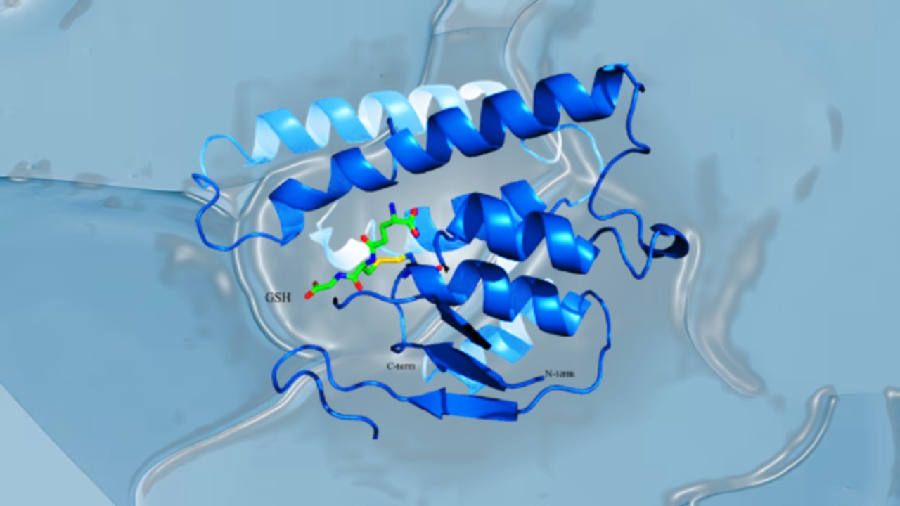
Resistance Mechanisms Unveiled: The Role of Glutathione S-Transferase in Cancer Therapy Failures
Understanding this dual role of GSTs as both protectors and accomplices to malignancies is central to tackling drug resistance.
Read More Articles
Myosin’s Molecular Toggle: How Dimerization of the Globular Tail Domain Controls the Motor Function of Myo5a
Myo5a exists in either an inhibited, triangulated rest or an extended, motile activation, each conformation dictated by the interplay between the GTD and its surroundings.
Designing Better Sugar Stoppers: Engineering Selective α-Glucosidase Inhibitors via Fragment-Based Dynamic Chemistry
One of the most pressing challenges in anti-diabetic therapy is reducing the unpleasant and often debilitating gastrointestinal side effects that accompany α-amylase inhibition.


