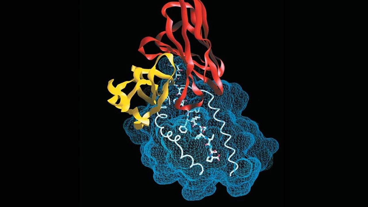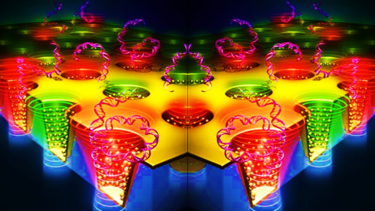
In the context of biology and medicine, nanotechnology encompasses materials, devices, and systems whose structure and function are relevant for small length scales, from nanometers (10−9 m) through microns.
Nanotechnology offers a number of advantages for biological research. In comparison to traditional molecular assays, nanoscale devices can differentiate at the level of single molecules and single cells. This great sensitivity can be used to characterise single-cell heterogeneity at extremely high throughput, revealing distinct hierarchies and subpopulations. This is owed to the microscopic structure of nanotechnologies which are typically comparable in size with biomolecules. As a result, they can travel more freely through the human body in comparison to larger molecules.
In addition to research, nanotechnology has contributed to a number of innovations across the drug development process including drug design and delivery. Nanotechnology is also evolving to deliver therapeutic agents to target sites.
Nanomedicine
Drug delivery systems
Polymeric nanomaterials are the ideal choice for efficient drug delivery. Polymeric refers to a large molecule made of smaller subunits. The high compatibility and biodegradability are a few of the properties that make polymeric nanomaterials desirable for delivering drugs that have poor solubility and low absorption rates. Nanospheres and nanocapsules are the two main categories of polymeric nanoparticles used as drug delivery systems.
Chitosan-based nanomaterials are widely used for continued drug release systems for various types of tissues including eye, intestinal and nasal regions. Chitosan is a naturally occurring by-product from the processing of shellfish.
One of the main advantages of using nano drug delivery systems is their ability to penetrate the tissue system. This facilitates easy uptake of the drug by cells, resulting in an efficient drug delivery at the specific target site. Furthermore, nanostructures stay in the blood circulatory system for a prolonged period and enable the release of drugs as per the specified dose. Therefore, they cause fewer systemic fluctuations, reducing the likelihood of adverse effects.
The method by which nanostructures deliver drugs is divided into two categories, passive and self-delivery. In passive delivery, drugs are integrated into the nanostructure via the hydrophobic effect. Hydrophobic refers to molecules that are repelled by water. The desired amount of drug will then be released at target sites due to the “low content of the drugs which is encapsulated in a hydrophobic environment”.
In passive drug delivery the drug carrier is transported systematically, and drawn to the target site by affinity influenced by properties like pH, temperature, molecular site and shape.
In self-delivery, the drugs are instead directly conjugated to the nano carrier. With this approach, the drug dissociates rapidly from the nanostructure, hence the timing of release is crucial to ensure the drug reaches the target site. If released prematurely, the bioactivity and efficacy will be significantly compromised.
With the active approach, drug targeting is facilitated by biological agents known as moieties e.g. antibodies and peptides. These agents are coupled with the nano drug delivery system to act as an anchor to receptor structures within the target site. The targets for drug delivery are primarily membrane-bound proteins including receptors, lipid structures or antigens on the cell surface.
Tissue engineering
In addition to drug development, nanotechnology has been used to create biocompatible scaffolds in the creation of implantable tissues. According to an article, these nanoscale structures have been engineered to resemble a native extracellular matrix which can control the release of drugs. In biology, an extracellular matrix is an intricate 3D network consisting of an array of extracellular macromolecules like proteins and carbohydrates. Also known as hard-tissue engineering, this nanotechnology is a relatively new concept used to engineer skeletal muscle tissue.
The mimicking of the native extracellular matrix is a crucial part of creating an optimum tissue microenvironment including appropriate mechanical strength, ease of monitoring cellular activities and delivering of bioactive agents require a nanoscale approach. Modified electrospinning is one method in which biological factors are incorporated into nanoscaffolds.
Gold and titanium nanoparticles have been used to enhance cellular functions like proliferation for bone and cardiac tissue regeneration. Several studies support the utility of gold nanoparticles especially as candidates for bone tissue regeneration. This particular group of nanoparticles have shown to influence osteoclast formation, while providing “protective effects on mitochondrial dysfunction in osteoblastic cells”. Osteoclasts are a type of bone cell that breaks down bone tissue.
These cells are critical for the maintenance of bone repair, metabolism and remodeling. Mitochondria function is critical for cellular function of which tissue regeneration cannot occur. Hence, protection of this component is a significant part of the success of bone regeneration seen so far.
Cardiac tissue regeneration has been a key focus for nano tissue engineering for many years. Human myocardium is a type of muscle tissue in the heart which typically fails to regenerate after tissue damage. This results in an insufficient number of cardiomyocyte cells and counteracting of scar tissue formation which leads to abnormal arrhythmia and often heart failure. Hence, this represents an unmet need for nanotechnology.
Engineered cardiac patches are considered a promising approach for regenerating the infarcted heart. Cardiac cells are implanted within the 3D nanoscaffolds, which provide the structural biochemical microenvironment. The formation of tissue arises from cell-cell and cell-matrix interactions facilitated by the scaffold structure. Once the tissue is engineered into a cardiac patch, it is attached to the scar tissue of the heart by a surgical operation, involving synthetic sutures or staples.
While cardiac patches have shown promising preclinical results, there are a significant number of challenges that need to be addressed before clinical implementation. Firstly, one of the issues with using metallic nanoparticles is the inability of these structures to be naturally broken down. As a result, the longer the period the structure remains in the body, the greater the likelihood of cytotoxic events. Secondly, the method by which the cardiac patches have been attached is using staples/sutures during open heart surgery. This is, of course, a highly invasive and risky process to which a less invasive alternative attachment method needs to be developed to reduce the risk of complications. 3D printing has been suggested as a recommendation for patch fabrication.
To be able to recapitulate the tissue microenvironment and functionality so well through nanotechnology represents a significant step forward in the successful regeneration of tissue. Despite significant setbacks with preclinical testing of cardiac tissue regeneration, research is ongoing to develop novel nano fabrications and less invasive delivery methods. Nanotechnology continues to play an important role across many therapeutic areas and drug development, and will continue to evolve in the next few years to solve current challenges in the field.
Charlotte Di Salvo, Former Editor & Chief Medical Writer
PharmaFEATURES
Subscribe
to get our
LATEST NEWS
Related Posts

Medicinal Chemistry & Pharmacology
Aerogel Pharmaceutics Reimagined: How Chitosan-Based Aerogels and Hybrid Computational Models Are Reshaping Nasal Drug Delivery Systems
Simulating with precision and formulating with insight, the future of pharmacology becomes not just predictive but programmable, one cell at a time.
Medicinal Chemistry & Pharmacology
Coprocessed for Compression: Reengineering Metformin Hydrochloride with Hydroxypropyl Cellulose via Coprecipitation for Direct Compression Enhancement
In manufacturing, minimizing granulation lines, drying tunnels, and multiple milling stages reduces equipment costs, process footprint, and energy consumption.

Medicinal Chemistry & Pharmacology
Decoding Molecular Libraries: Error-Resilient Sequencing Analysis and Multidimensional Pattern Recognition
tagFinder exemplifies the convergence of computational innovation and chemical biology, offering a robust framework to navigate the complexities of DNA-encoded science


