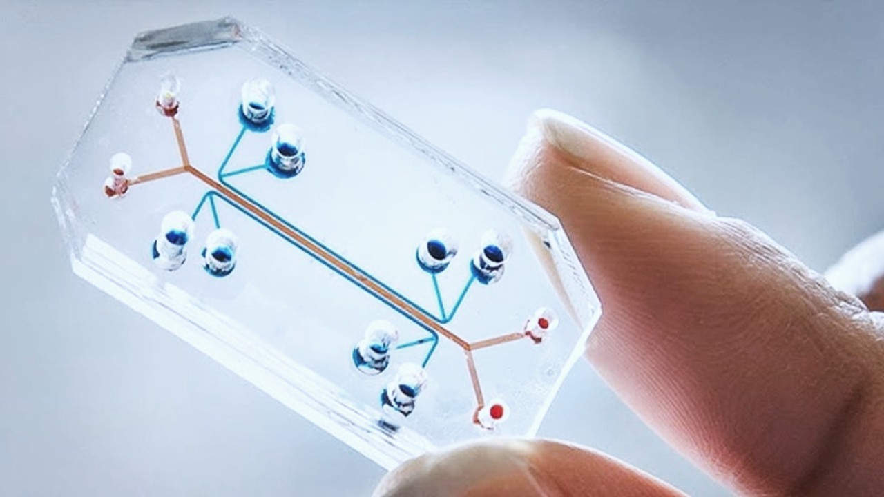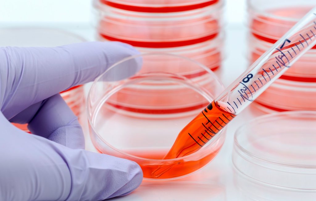
Preclinical studies are an integral part of drug development in terms of screening drug candidates for therapeutic efficacy and potential toxic events. 3D culture and organ-on-chip technology are a few examples of the innovations in in vitro/in vivo modelling to improve the representation of human physiology for preclinical testing.
3D culture
Developments in cell cultures have arisen from the need for “new preclinical models that better recapitulate in vivo biology and microenvironmental factors”. This is a key area in drug development – the better the preclinical models, the more opportunities to address the challenges of drug discovery and drug repurposing. 3D cultures are an example of the latest advancements in cell-based assays. In comparison to 2D cultures which are performed in vitro, 3D cell culture occurs within the cell’s native environment, known as in vivo.
The purpose of cell-based assays is to accurately model the cellular environment of an organism/target tissue in order to test the safety and efficacy of a potential drug. 2D cultures are simple, low-cost assays that have served the industry well for many years. However, there are a number of limitations to this method. Firstly, “2D cultured cells do not mimic the natural structures of tissues or tumours”.
Due to the process of isolating cells from native tissue and transferring to 2D conditions, the morphology of cells is disturbed. Hence, this results in alterations to the cellular interactions with the external environment. Cell-cell and cell-extracellular environment interactions are a key part of tissue physiology, responsible for cell differentiation, drug metabolism, and a number of other cellular functions. Therefore, if a preclinical model fails to accurately represent native tissue, the pharmacokinetics of drug candidates may not be reliable in terms of prediciting pharmacokinetics in clinical trials.
3D cell cultures grow cells into 3D aggregates/spheroids using a scaffold/matrix in a scaffold-free manner. It is this spheroid structure that is suggested to more accurately represent the native microenvironment and the cellular interactions. Furthermore, the third dimension has been suggested to be a crucial feature in understanding the influence of the “spatial organization of the cell surface receptors engaged in interactions with surrounding cells”. The 3D structure better facilitates the signal transduction and consequently influences cell behaviour which represents cell response more similar to in vivo compared to 2D culture.
3D cell cultures have served as a model for a variety of experimental oncology studies using radiotherapy, chemotherapy, and immunotherapy. Due to the growth of solid tumours in 3D, the spatial arrangement of cells in the structure determines the level of interaction between each of them. Hence, 3D cell cultures are suggested to better mimic the natural environment of solid tumours than the 2D monolayer cell cultures.
A 2020 study investigated the application of 3D cell cultures in preclinical testing for biomarker discovery and validation. The team investigated environment-dependent miRNA expression changes in colorectal adenocarcinoma (CRC) using next-generation sequencing technology.
It was revealed that the dysregulation of miRNA expression is dependent on the 3D microenvironment, and potentially “triggers essential molecular mechanisms predominantly including the regulation of cell adhesion…important in CRC initiation and development”. This is an important step forward in gaining understanding of the molecular mechanism of CRC and potentially further studies assessing the implication of miRNA signature in CRC.
Organ-on-a-chip technology
According to a 2020 article, organ-on-a-chip technology (OOAC) is on the list of top ten emerging technologies and refers to a physiological organ biomimetic system built on a microfluidic chip. OOAC is a biomimetic model which refers to a platform that simulates the tissue microenvironment and mechanical stimulation of a human organ. In addition, the microfluidic chip can regulate key parameters including concentration gradients, cell patterning, and mechanical stimulation like shear force.
Reflecting the structural and functional characteristics of human tissue continues to be an obstacle in preclinical research. While animal models have been useful as in vitro models for drug discovery, they are limited by species-exclusive physiology. OOAC is an important asset that can potentially be used to predict response to an array of stimuli including drug responses and environmental effects in human disease.
There are four key components to OOAC: microfluidics, living cell tissues, stimulation or drug delivery, and sensing. Microfluidics refers to the manipulation and processing of microscale fluids. Microfluidics is used to deliver target cells to a “pre-designated location” in addition to forming fluid input/waste output during cell culturing. The living cell tissue refers to “components that spatially align a particular cell type in the case of 2D or 3D systems”.
While 3D cell cultures can more accurately simulate the in vivo microenvironment, 2D cultures are often chosen as a more cost-effective approach to cell cultures. The sensing component refers to a “transparent chip based visual function evaluation system”. This embedded component acts to assess the overall functionality of the model.
The first example of an automated organ-on-a-chip was seen in a 2019 study investigating the “automated microfluidic cell culture of stem cell derived dopaminergic neurons”. Dopaminergic neurons are a type of nerve cell in the brain susceptible to damage in Parkinson’s. The study constructed an automated cell culture system to enable the monitoring of patient-derived Parkinson’s human neuroepithelial stem cells. In comparison to manual cell cultures which are subject to human error, an automated system uses a liquid handler and a robot to move receptacles.
The success of the automated system in maintaining the neuroepithelial cells was supported by calcium imaging which demonstrated spontaneously filing neurons as well as immunostaining for neuronal markers. According to the team, the 3D automated cell model demonstrates a “proof-of-concept for automated generation of personalised in vitro neuronal models from human neuroepithelial stem cells via a microfluidic cell culture approach”.
The ultimate goal is to integrate numerous organs onto a single chip, to build a more complex multi-organ chip model.This could open the door to achieving a “human-on-a-chip”. A human-on-a-chip would support drug development in terms of studying drug pharmacokinetics on a whole system level, optimising drug efficacy and minimising adverse effects.
Charlotte Di Salvo, Former Editor & Chief Medical Writer
PharmaFEATURES
Subscribe
to get our
LATEST NEWS
Related Posts
Molecular Biology & Biotechnology
Myosin’s Molecular Toggle: How Dimerization of the Globular Tail Domain Controls the Motor Function of Myo5a
Myo5a exists in either an inhibited, triangulated rest or an extended, motile activation, each conformation dictated by the interplay between the GTD and its surroundings.
Drug Discovery Biology
Unlocking GPCR Mysteries: How Surface Plasmon Resonance Fragment Screening Revolutionizes Drug Discovery for Membrane Proteins
Surface plasmon resonance has emerged as a cornerstone of fragment-based drug discovery, particularly for GPCRs.
Read More Articles
Designing Better Sugar Stoppers: Engineering Selective α-Glucosidase Inhibitors via Fragment-Based Dynamic Chemistry
One of the most pressing challenges in anti-diabetic therapy is reducing the unpleasant and often debilitating gastrointestinal side effects that accompany α-amylase inhibition.


