The world of drug discovery is evolving. Once rooted in conventional methods that primarily relied on animal models and chemical screening, it is now shifting toward a more refined approach, integrating human biology at the earliest stages of development. The limitations of traditional drug discovery methods, while offering insights, have led to a growing demand for more accurate and predictive models. As we progress toward a future that embraces personalized medicine, understanding the transformation from traditional preclinical models to innovative human tissue-based approaches is paramount. This shift holds the potential to bridge the gap between preclinical testing and clinical success, reducing the notorious drug attrition rates that have long plagued pharmaceutical research.
Rethinking Cancer Models: A Closer Look at Human Malignancies
The development of preclinical cancer models has historically centered around reproducing the genetic and physiological aspects of human malignancies in vitro and in vivo. Traditional methods such as immortalized cell lines and genetically engineered mouse (GEM) models have provided valuable insights, but they fall short in fully representing the complexities of human cancer. Cancer progression is driven not only by intrinsic genetic mutations but also by external factors, including interactions with the immune system and metastatic potential.
In vitro systems, like high-throughput screening (HTS) platforms using immortalized cell lines, target specific genetic pathways but miss out on capturing the multi-faceted nature of tumor growth and immune evasion. On the other hand, GEM models are engineered to carry human genetic mutations in immunocompetent mice, providing a more realistic scenario. However, even with advancements like CRISPR/Cas9 for genetic editing, these models are inherently limited by species differences.
The need for humanized models is apparent, especially when considering the role of the immune system in cancer progression. The traditional use of human tissue xenografts in immunocompromised mice, though helpful for studying tumor formation and drug efficacy, lacks the critical component of an intact immune system. A shift toward human tissue-based models could enhance our ability to predict therapeutic outcomes, particularly in the burgeoning field of immuno-oncology.
Tackling Obesity and Metabolic Diseases: Beyond the Mouse Model
The rising prevalence of obesity and its associated metabolic disorders, such as type-2 diabetes and non-alcoholic fatty liver disease, underscores the urgency of developing better preclinical models. Current models predominantly use genetically or diet-induced obesity in small animals like mice. While these models offer a controlled and reproducible system for studying metabolic dysfunction, they fail to capture the intricacies of human metabolism and immune response.
One key limitation lies in the differences between human and mouse adipose tissue. In humans, visceral fat depots, particularly omental and mesenteric fat, play a central role in obesity-related complications. In contrast, mice have relatively small visceral fat depots and predominantly store fat in epididymal regions, which do not correspond to human obesity patterns. This disparity affects the study of adipose tissue inflammation, insulin resistance, and endocrine dysfunction—all hallmarks of metabolic disease.
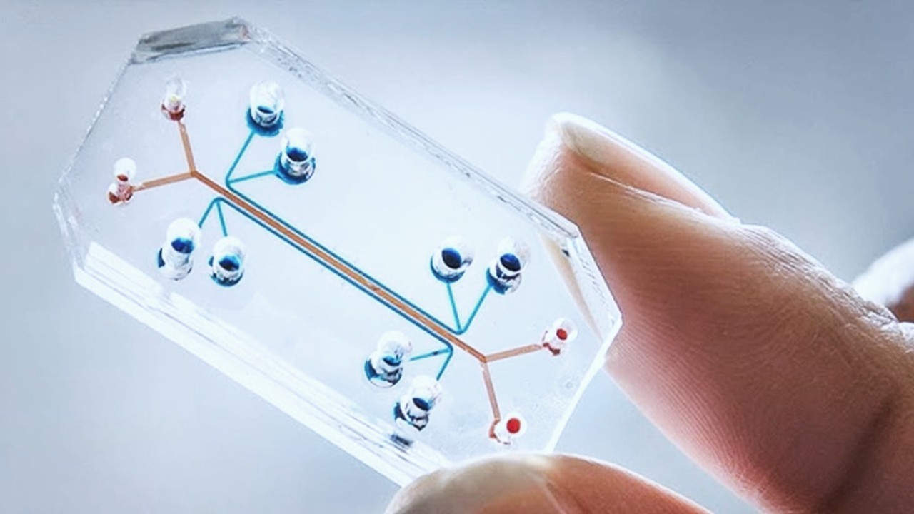
Innovative approaches using human adipocyte cultures and co-culture systems that incorporate immune cells could provide a more physiologically relevant platform for studying obesity and its comorbidities. These systems would enable researchers to investigate the complex interactions between adipose tissue, the immune system, and metabolic pathways, offering new avenues for drug discovery in this critical area of global health.
The Vaccine Development Dilemma: Human Models for a Global Need
The COVID-19 pandemic brought vaccine development to the forefront of public consciousness, highlighting the challenges in creating effective vaccines. Traditional vaccine development relies heavily on small and large animal models, which often fail to replicate the human immune response accurately. The immunological differences between humans and animals, particularly in the complex cascade of humoral immunity, pose significant barriers to developing effective vaccines.
Non-human primates are commonly used to bridge this gap, but species-specific differences in immune regulation and pathogen response still hinder progress. For instance, certain pathogens have restricted tropisms, meaning they only infect specific species. This limitation makes it difficult to model human immune responses to these pathogens in animals. Moreover, the safety concerns associated with vaccines—such as the potential for immune enhancement, where a vaccine exacerbates disease upon exposure to the pathogen—further complicate the development process.
Human cell-based models hold promise in addressing these challenges. Co-culture systems of human immune cells can offer more accurate predictions of vaccine efficacy and safety, particularly for respiratory viruses like RSV, where traditional animal models have fallen short. As vaccine development expands to include emerging areas like cancer immunotherapy, the demand for humanized models that can faithfully replicate immune responses will only increase.
Understanding Human Development: A New Frontier in Disease Modeling
Developmental disorders affect millions of live births globally, with profound impacts on public health and healthcare costs. However, the study of human development in preclinical models remains a significant challenge due to the ethical and practical limitations of accessing human fetal tissues. As a result, traditional animal models have been used to study teratogenic effects—the harmful impacts of drugs on fetal development. These models, while informative, do not fully capture the nuances of human development.
Thalidomide, a notorious example of drug-induced birth defects, underscores the importance of accurate preclinical models for detecting teratogenic risks. Despite being deemed safe in animal studies, thalidomide caused severe limb deformities in thousands of children before it was withdrawn from the market. This tragedy highlights the limitations of relying on animal models to predict human developmental outcomes.
Innovative models that use human pluripotent stem cells to recreate early developmental stages could revolutionize the study of developmental disorders and teratogenic risks. By using these models to screen for potential drugs or toxic compounds, researchers could identify harmful effects before they reach clinical trials, preventing future tragedies and advancing our understanding of human development.
Degenerative Diseases: Modeling Human Aging and Neurodegeneration
The global burden of neurodegenerative diseases like Alzheimer’s, Parkinson’s, and multiple sclerosis is growing, driven by aging populations. Despite significant efforts to develop treatments, progress has been slow, in part due to the limitations of traditional preclinical models. Neurodegenerative diseases are often complex and involve multiple cell types and non-cell-autonomous mechanisms, where the failure of one cell type triggers dysfunction in others.
Current mouse models, such as those used to study Alzheimer’s, replicate some aspects of the disease, like the formation of amyloid plaques. However, they fail to capture other critical features, such as the tau protein tangles that are characteristic of the later stages of human Alzheimer’s disease. This limitation hampers the development of therapies that target these advanced stages of the disease.
Human-based models, particularly those that incorporate multiple cell types and mimic the blood-brain barrier, offer a more promising platform for studying neurodegenerative diseases. These models could provide insights into disease mechanisms and drug transport issues, helping to overcome the challenges of developing effective treatments for these complex disorders.
Toward a New Era of Drug Discovery
The shift from traditional preclinical models to human tissue-based systems represents a fundamental change in how we approach drug discovery. By incorporating human biology at the earliest stages of development, these innovative models hold the potential to transform the landscape of medicine, offering more predictive and accurate insights into disease mechanisms and therapeutic efficacy. As we move toward a future where personalized medicine becomes the norm, the development of human-relevant preclinical models will be critical in bridging the gap between bench and bedside.
Engr. Dex Marco Tiu Guibelondo, B.Sc. Pharm, R.Ph., B.Sc. CpE
Editor-in-Chief, PharmaFEATURES

Subscribe
to get our
LATEST NEWS
Related Posts
Molecular Biology & Biotechnology
Myosin’s Molecular Toggle: How Dimerization of the Globular Tail Domain Controls the Motor Function of Myo5a
Myo5a exists in either an inhibited, triangulated rest or an extended, motile activation, each conformation dictated by the interplay between the GTD and its surroundings.

Drug Discovery Biology
Unlocking GPCR Mysteries: How Surface Plasmon Resonance Fragment Screening Revolutionizes Drug Discovery for Membrane Proteins
Surface plasmon resonance has emerged as a cornerstone of fragment-based drug discovery, particularly for GPCRs.
Read More Articles
Designing Better Sugar Stoppers: Engineering Selective α-Glucosidase Inhibitors via Fragment-Based Dynamic Chemistry
One of the most pressing challenges in anti-diabetic therapy is reducing the unpleasant and often debilitating gastrointestinal side effects that accompany α-amylase inhibition.


