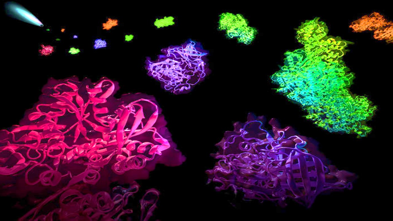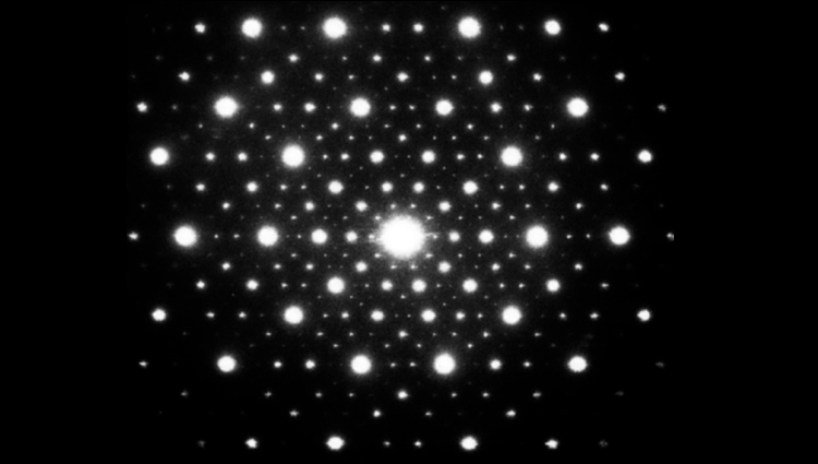
Microcrystal electron diffraction (microED) has the potential to accelerate areas of drug development like drug discovery, rivalling conventional techniques, like x-ray crystallography. Recent studies have demonstrated how microED can determine the 3D structure of molecules at a higher resolution and greater speed, but is not limited by the small size of crystal samples. Despite the slow adoption by the pharma industry, microED continues to emerge in an increasing number of studies for small molecule drug design.
Introduction
MicroED is an emerging technique in structural biology that enables the study of biomolecules from the micrometer-sized crystals that are too small for the conventional x-ray crystallography. microED allows fast, high-resolution 3D structure composites of small chemical compounds and biological macromolecules.
The data acquired by microED is acquired via a cryo-transmission electron microscope. The crystallisation process is very similar to that of x-ray crystallography, however much smaller crystals can be analysed because the interaction with electrons is stronger than it is with x-rays.
X-ray crystallography continues to remain the gold standard for determining molecular structure (via crystallisation) for now, but is limited by a bottleneck problem, as it requires the generation of large, well-ordered crystals. In addition, the unpredictability of the protein crystallisation process can be a challenge, as it remains a ‘trial and error’ based procedure, which can make it time-consuming and resource heavy.
More recently, microED has been emerging as a popular choice for determining the structure of molecules like proteins, peptides and small organic molecules. While x-ray crystallography continues to be refined, some pharma companies are looking towards microED as a potential alternative which can provide results at higher resolutions of molecular structure at an atomic level.
How might it be useful for drug development?
The first application of MicroED to the study of peptides was back in 2015, which determined the novel structure of a biomolecule for the first time. The structure was a segment of the protein involved in the pathology of Parkinson’s, known as alpha-synuclein. Techniques like X-ray free-electron lasers were unable to determine the structure of the microscopic crystal structure. MicroED however, was able to facilitate the structure determination of these peptides in a rapid timeframe.
Understanding the crystalline structure of small molecules is of paramount importance in small molecule drug development. The crystal form of an active pharmaceutical ingredient (API) and the interactions that hold it together can have important consequences on a number of properties like “stability, tableting properties, solubility and dissolution rates, ultimately affecting bioavailability, potency, and even toxicity”.
It is therefore desirable to determine the 3D crystal structure of an API in order to engineer the optimal form for development. The reason why researchers are beginning to look towards microED is that it can work with crystal structures of almost any size, in comparison with x-ray crystallography which requires larger crystals.
MicroED has evolved significantly over the past few years thanks to the integration of new technologies. The computerisation of transmission electron microscopes for example, has allowed software to be developed that can semi-automatically collect 3D data in less than an hour.
In a recent article, a team of researchers at NanoImaging Services (UK) emphasised that using microED would be particularly useful in drug development when dealing with small molecules, many of which are in small sample quantities in the drug discovery phase. It was stated that “recent publications already show how it could help to speed up the development of new drugs, and we are eagerly anticipating how it might impact the volume and breadth of data we are able to share”.
One of the greatest advantages for using microED is that users can directly visualise the nano-/microcrystals and choose the best ones to collect data from. Even the nanocrystals can produce high-quality and high-resolution data, which can be used for small molecule structure determination.
Visualising drug binding interactions
A 2020 study published in Nature recently demonstrated the progression of using microED in drug development so far, investigating its role in structure-based drug design. In this study, it was demonstrated that microED can effectively be used to visualise protein-inhibitor-binding interactions, by determining the structure of a molecule known as HCA in complex with a clinical drug.
Two of the main advantages for using this method was that less sample material was required and faster diffusion of the ligand was facilitated. It was also suggested that microED could be used to complement existing methods like x-ray crystallography when working with larger crystal structures for better resolution.
Visualising the ligand binding interactions is an integral part of structure-based drug design as it demonstrates the structural knowledge of the protein-active site and the molecular interactions of small molecule binding. In the case of designing drugs which inhibit the function of a protein, it is essential to understand the structure of the active site and ligand interaction in order to assess whether a drug candidate is structurally suitable to bind/interact with the protein.
Challenges
Of course, the integration of emerging technologies and techniques doesn’t come without challenges.
The adoption of microED in medicinal chemistry has been slow due to factors like the steep requirements for instrumentation, infrastructure and expertise. These key factors are taken into account when pharma companies assess whether they can justify the capital investments, especially so if only a small group of projects would benefit.
It has been suggested that improving the access to service facilities, the speed and automation of data collection and processing will make small molecule microED more broadly accessible, especially for R&D in the pharmaceutical industry.
In terms of future perspectives, the studies so far show how microED could benefit drug discovery significantly. High-throughput approaches and automated collection of microED data may be the next step to increase the amount of data collected in a single session and the speed of the overall workflow.
Charlotte Di Salvo, Lead Medical Writer
PharmaFeatures
Subscribe
to get our
LATEST NEWS
Related Posts
Molecular Biology & Biotechnology
Myosin’s Molecular Toggle: How Dimerization of the Globular Tail Domain Controls the Motor Function of Myo5a
Myo5a exists in either an inhibited, triangulated rest or an extended, motile activation, each conformation dictated by the interplay between the GTD and its surroundings.
Medicinal Chemistry & Pharmacology
Invisible Couriers: How Lab-on-Chip Technologies Are Rewriting the Future of Disease Diagnosis
The shift from benchtop Western blots to on-chip, real-time protein detection represents more than just technical progress—it is a shift in epistemology.

Medicinal Chemistry & Pharmacology
Designing Better Sugar Stoppers: Engineering Selective α-Glucosidase Inhibitors via Fragment-Based Dynamic Chemistry
One of the most pressing challenges in anti-diabetic therapy is reducing the unpleasant and often debilitating gastrointestinal side effects that accompany α-amylase inhibition.


