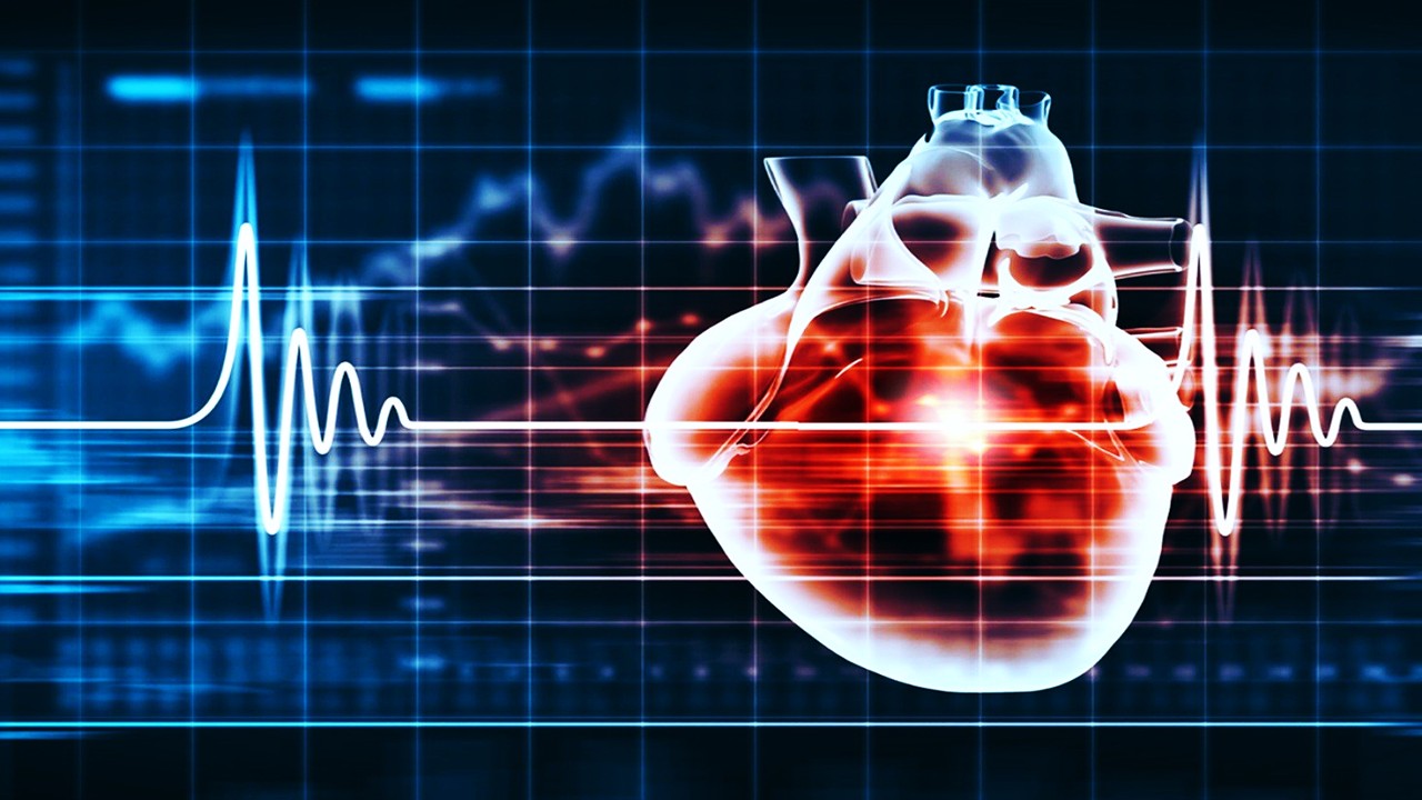
The Silent Signalers of the Human Body
Every cell in the human body participates in an elegant, ceaseless dialogue, and some of the most critical messengers are entirely invisible to the naked eye. Encased in lipid bilayers and coursing through our blood, urine, saliva, and cerebrospinal fluid are extracellular vesicles—minute parcels released by virtually every cell type. Among these, exosomes and microvesicles stand out for their ability to transport biomolecules like proteins, lipids, mRNA, and even double-stranded DNA. These are not mere cellular waste products; they are functional emissaries of physiological and pathological states, rich in biological information reflective of their parent cells.
Exosomes typically span 30 to 100 nanometers and are formed by inward budding within endosomal compartments before being released through multivesicular body fusion. Microvesicles, larger and more heterogeneous in size—ranging from 100 to 1000 nanometers—are generated by outward budding of the plasma membrane. While the morphological distinctions are subtle, their biogenic divergence is significant, influencing their molecular payload and interaction mechanisms. Yet, despite their ubiquitous presence and molecular richness, the clinical community has struggled to harness their diagnostic power.
Part of the challenge lies in the biochemical indistinguishability between these vesicle subtypes. Both can share surface markers and coexist within the same biological fluids, complicating attempts to isolate them with precision. For diseases where early molecular signals are faint—such as neurodegenerative disorders or early-stage cancers—this specificity is not a luxury, but a necessity. Traditional techniques such as ultracentrifugation and polymer-based precipitation have proven inadequate for scalable, pure, and rapid vesicle isolation.
Emerging lab-on-chip (LOC) technologies, leveraging principles of microfluidics, surface chemistry, and physics, are poised to redefine how these vesicles are captured, sorted, and studied. LOC systems are engineered miniaturized laboratories, compressing complex diagnostic workflows into palm-sized cartridges that deliver clinical-grade precision. They promise not just a technical upgrade, but a conceptual revolution: shifting diagnostics from centralized labs to decentralized, point-of-care settings.
What makes this transition transformative is not merely size reduction. It’s the convergence of automation, specificity, and integrative modularity—each tailored to the biological quirks of exosomes and microvesicles. With these technologies, researchers are not just finding biomarkers; they are listening to the molecular whispers of disease before it screams.
Microfluidic Sculptures: Engineering Biological Selectivity
The architecture of LOC platforms is as much an exercise in physics as in biology. Microfluidics, the underlying engine of these systems, channels minuscule fluid volumes through microscale pathways with unprecedented control. Within these channels, passive and active sorting techniques exploit the vesicles’ biophysical properties to isolate them from biological chaos. These include immunoaffinity-based strategies that latch onto vesicle-specific markers and size-exclusion membranes that act as molecular sieves.
Passive sorting modules are often the starting point. Immunoaffinity techniques immobilize antibodies specific to exosomal surface proteins like CD63, CD81, or CD9 along microchannel walls. When a patient’s serum flows through, exosomes displaying these antigens are captured in situ. One such innovation, the Exochip, employs a serpentine channel architecture lined with CD63 antibodies to selectively trap cancer-derived exosomes, enabling downstream RNA analysis. This level of biological targeting turns a microchannel into a molecular gatekeeper, filtering out noise while retaining signal.
Size-exclusion modules provide an orthogonal layer of selectivity. By integrating nanomembranes with predefined pore sizes into fluidic networks, researchers can exclude larger vesicles or protein aggregates, isolating exosomes based on their diminutive dimensions. Platforms like ExoTIC and Exodisc use dual-membrane filtration to enrich exosomal populations with remarkable throughput, often outperforming ultracentrifugation in purity and yield. The precision is so fine-tuned that exosomes can be retained while free nucleic acids and protein contaminants are flushed out.
More dynamic are flow-induced systems, where the interplay of fluid inertia, pressure gradients, and viscoelastic forces guides vesicles into predetermined streamlines. Deterministic lateral displacement (DLD) chips, for instance, sort vesicles by size using pillar arrays that physically divert their path. Meanwhile, viscoelastic microfluidics, leveraging polymers like poly(oxyethylene), sort vesicles based on size-induced lift forces within laminar flows. These devices achieve continuous, label-free, and highly selective exosome isolation at clinically relevant throughputs.
Despite their elegance, these systems are not without limitations. Immunoaffinity-based capture is constrained by surface marker variability; size-exclusion risks clogging; and flow-based systems require precise physical modeling to function optimally. But their modular nature makes them ideal building blocks—each addressing a different facet of the exosome isolation conundrum.
Electricity, Magnets, and Sound: The Physics of Precision Isolation
Beyond passive systems, active LOC modules introduce external forces to elevate vesicle sorting from efficient to exquisite. These platforms deploy electric fields, magnetic gradients, or acoustic waves to manipulate vesicles according to their dielectric, magnetic, or mechanical properties. What emerges is a multidimensional toolkit capable of isolating vesicles invisible to traditional methods—not by size or antigen alone, but by how they interact with the physical world.
Electrokinetic platforms leverage dielectrophoresis, where non-uniform electric fields generate forces that attract or repel vesicles based on their dielectric constants. This allows for label-free, real-time sorting with minimal thermal damage. In one striking example, glioblastoma-derived exosomes were separated from plasma using an AC field and patterned electrodes, enabling sub-30-minute isolation. The process capitalized on the unique electrical signatures of cancer-derived vesicles, offering a new axis of diagnostic specificity.
Magnetophoretic methods build on similar principles but utilize superparamagnetic beads conjugated with capture ligands. Once the vesicles bind to these beads, an external magnetic field extracts the vesicle-bead complexes from a flowing sample. This approach shines in scalability and biocompatibility. Tools like the ExoSearch platform integrate immunomagnetic capture with downstream multiplexed biomarker analysis, detecting tumor markers in ovarian cancer plasma with a clinical finesse comparable to ELISA.
Acoustofluidics, the third pillar, manipulates vesicles through bulk or surface acoustic waves. SAW-based platforms, particularly those using piezoelectric substrates, can separate exosomes from larger EVs or cellular debris with contactless precision. In a dual-module acoustofluidic device, the first wavefield eliminates cells and debris while the second isolates exosomes—achieving a purity level previously only possible with hours of centrifugation. Crucially, these methods preserve vesicle integrity, a vital consideration for downstream molecular assays.
Each of these active platforms opens diagnostic frontiers that were, until recently, inaccessible. They allow researchers to not only capture exosomes of interest, but to dissect vesicle subpopulations with surgical accuracy. The physics is nontrivial, but the clinical implication is profound: precision diagnostics, governed not just by biology, but by the mastery of force.
From Counting to Profiling: On-Chip Analytical Modules
Capturing extracellular vesicles is only the beginning. For true clinical utility, their enumeration and molecular characterization must follow—ideally within the same device. Modern LOC platforms are now incorporating modules that quantify and analyze vesicles in real time, transforming raw biological material into interpretable data.
Counting vesicles begins with adapting flow cytometry principles into miniaturized formats. Microflow cytometers, outfitted with silica capillaries and optical fibers, have achieved throughputs rivaling their benchtop counterparts. Innovations in fluorescent tagging of surface markers allow for subtype discrimination down to 50 nanometers, enabling phenotypic profiling of exosomes by origin or disease association. These advancements have transformed exosome enumeration into a high-resolution, digital metric.
Complementing optical methods are electrical detection strategies. Devices built around split-ring resonators and field-effect transistors detect shifts in dielectric environments as vesicles transit through microchannels. When optimized, these systems can detect the passage of individual vesicles, providing real-time concentration data with label-free specificity. The electric signals are processed and interpreted in milliseconds, opening possibilities for continuous monitoring applications.
For labs prioritizing hybridization, integrated systems now combine optical and electrical modalities. These platforms utilize fluorescence for surface marker detection while simultaneously measuring impedance changes. Such dual-readout systems are not just redundant—they offer orthogonal validation, boosting diagnostic reliability. When rare subpopulations of vesicles must be detected, as in early-stage cancer or neurodegeneration, this redundancy becomes a strength.
A critical consideration in enumeration is assay sensitivity. Too often, standard cytometry underestimates vesicle counts due to size limitations or signal noise. LOC platforms, with their customized geometries and engineered flow environments, can overcome these biases. Their compact form factors also allow for multiplexed analysis—multiple markers, multiple vesicle types, all within a single cartridge.
The promise of on-chip analysis is clear: democratized, decentralized diagnostics that deliver complex biological readouts without the need for centralized laboratories. And at the heart of this capability lies an elegant synergy of microengineering and biomolecular science.
Opening the Vesicular Vault: Lysis and Intravesicular Discovery
Once isolated and counted, exosomes and microvesicles must be breached—carefully—to reveal their inner cargo. This cargo, composed of nucleic acids, proteins, and lipids, is what transforms these vesicles from simple biomarkers into molecular portraits of their origin. However, lysing vesicles without damaging their sensitive contents remains a technical high-wire act. Lab-on-chip platforms now include meticulously engineered lysis modules, designed not to destroy but to unlock.
Chemical lysis remains one of the simplest approaches. Surfactants like Triton X-100 or SDS rupture vesicle membranes through solubilization, releasing internal contents. This method is often embedded within microfluidic chips via preloaded buffers and microwell architectures, allowing precise spatiotemporal control over the reaction. But the chemical route is slow and sometimes incompatible with downstream assays—DNA degradation and enzyme interference are frequent concerns. Yet, its universality makes it a foundational module in many integrated systems.
In contrast, physical lysis methods leverage microfabricated electrodes or acoustic fields to induce membrane rupture. Electric-field lysis, or electroporation, exploits differences in transmembrane potential to produce nanoscale pores, through which internal cargo escapes. When applied within microchannels with tightly regulated field gradients, this technique allows high-throughput lysis with minimal denaturation. The result is a clean sample, enriched in RNA or protein, ready for real-time amplification or detection.
More novel is the application of surface acoustic waves (SAWs). Here, interdigitated transducers on piezoelectric substrates generate mechanical perturbations that shear open vesicles without the need for detergents. The electric field and mechanical agitation act synergistically, enabling lysis of even the smallest exosomes. In a notable application, pancreatic cancer-derived exosomes were lysed within 30 seconds using a SAW microfluidic module, releasing miRNA for downstream quantification. This rapid, contactless method preserves molecular integrity better than most conventional techniques.
As the demand grows for high-fidelity exosome analysis, hybrid approaches have emerged. These systems combine initial capture with SAW-based lysis followed by electric-field-driven nucleic acid extraction—seamlessly integrated within one fluidic path. This degree of orchestration was once unimaginable in traditional lab workflows but is now routine in advanced LOC platforms. It represents a paradigm shift not just in scale but in conceptual elegance.
Ultimately, lysis is no longer an end-point procedure—it’s the gateway to molecular profiling. And the way this gateway is engineered can mean the difference between a diagnostic breakthrough and a signal lost in biological noise.
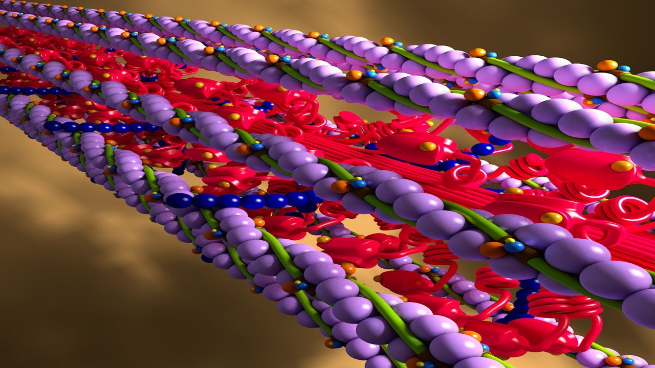
Sequencing the Whisper: On-Chip Nucleic Acid Detection
Extracellular vesicles do not simply transport genetic material—they encode stories of cellular fate, dysfunction, and resilience. Their cargo of miRNAs, mRNAs, and even oncogenic DNA fragments represents a trove of clinical insights. Unlocking this information requires highly sensitive, integrable nucleic acid detection systems—ideally within the same microfluidic architecture that isolates and lyses the vesicles.
Polymerase Chain Reaction (PCR) remains the gold standard for amplification and detection, but miniaturizing this process onto a chip demands radical engineering. Isothermal amplification techniques like loop-mediated amplification (LAMP) have been favored for their thermal simplicity. These methods function at constant temperatures, removing the need for complex thermal cycling hardware. When embedded within transparent PDMS chips, and paired with low-cost CMOS cameras for fluorescent readouts, the result is a portable diagnostic laboratory that fits in your palm.
In advanced systems, magnetic bead-based nucleic acid capture is used to purify genetic material before amplification. Droplet magnetofluidics, for instance, manipulate charged magnetic particles across discrete reagent chambers—binding nucleic acids, washing them, and delivering them to amplification wells. One such system achieved Hepatitis C viral RNA detection in clinical serum samples with sensitivity rivaling centralized hospital tests, all within a disposable cartridge.
Electrochemical strategies provide an alternative to optical systems, enabling label-free detection with sub-picomolar sensitivity. These methods often rely on hybridization chain reaction (HCR) systems, where target miRNAs induce a self-assembling cascade of DNA hairpins. The resulting amplified DNA structures change the local charge environment at an electrode, producing a quantifiable impedance signal. These systems are remarkably stable, inexpensive, and integrable into mass-manufacturable chips.
More futuristic are nanoscale field-effect transistors (FETs) and DNA arrays embedded within fluidic channels. These sensors detect DNA hybridization events in real time, converting the biorecognition event into a discrete electrical signal. Some FET-based platforms now detect sequence mismatches as subtle as two nucleotides, offering mutation specificity without requiring PCR. These technologies hold particular promise for detecting tumor-specific mutations from exosomal DNA in blood.
By embedding these tools within LOC systems, nucleic acid analysis becomes rapid, decentralized, and automated. The implications for infectious disease detection, cancer monitoring, and precision therapeutics are staggering. What once required a clinical lab and hours of hands-on work now happens invisibly—inside the translucent walls of a microfluidic chip.
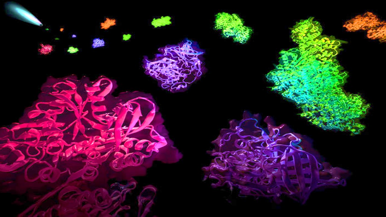
Decoding the Proteomic Language of Vesicles
If nucleic acids are the script, then proteins are the performance—revealing not just potential but phenotype. Exosomal and microvesicular proteins, encapsulated during vesicle formation, reflect the functional state of the parent cell. Detecting these proteins with fidelity and speed is key to transforming vesicle analysis into clinical practice. LOC platforms are now rising to this challenge with elegant proteomic modules designed for both specificity and scalability.
Fluorescence-based detection remains the dominant strategy. Recent platforms integrate photonic crystals to amplify the signal from standard ELISA-like assays, achieving sub-picogram-per-milliliter detection of cytokines like IL-3 and TNF-α. This amplification is not merely optical—it’s diagnostic, enabling the detection of immune shifts or tumor markers with clinical resolution from minute sample volumes. These tools compress the entire ELISA workflow into a single fluidic path, reducing both hands-on time and reagent cost.
Label-free strategies are gaining traction for their simplicity and low consumable burden. Electrochemical impedance spectroscopy and cyclic voltammetry, deployed across self-assembled monolayers of gold electrodes, can quantify antigen–antibody interactions with remarkable precision. These systems respond to subtle changes in the local electrochemical environment, eliminating the need for secondary antibodies or fluorescent dyes. One recent device achieved zeptomolar sensitivity in Alzheimer’s diagnostics using prion-like protein probes.
Nanomaterials further enhance detection capabilities. Graphene, molybdenum disulfide, and other 2D materials offer high surface-area-to-volume ratios and exceptional electron mobility—ideal for building ultrasensitive biosensors. When functionalized with capture ligands, these materials convert minute protein binding events into measurable current changes. Such systems have already demonstrated femtomolar detection of breast and prostate cancer markers in clinical fluids.
Multiplexing is the next frontier. LOC platforms are beginning to support arrays of sensors that can simultaneously detect dozens of exosomal proteins. This opens the door to signature-based diagnostics—using complex proteomic fingerprints rather than single analytes. In inflammatory or autoimmune conditions where multiple cytokines change simultaneously, this holistic view provides a richer, more actionable clinical picture.
The shift from benchtop Western blots to on-chip, real-time protein detection represents more than just technical progress—it is a shift in epistemology. It moves proteomics from post-hoc analysis to point-of-care insight, from hypothesis-driven to discovery-enabled. In the hands of clinicians, it becomes not just a diagnostic tool, but a lens into the molecular theater of disease.
Study DOI: https://doi.org/10.3390/s18103175
Engr. Dex Marco Tiu Guibelondo, B.Sc. Pharm, R.Ph., B.Sc. CpE
Editor-in-Chief, PharmaFEATURES

Subscribe
to get our
LATEST NEWS


Related Posts

Medicinal Chemistry & Pharmacology
Designing Better Sugar Stoppers: Engineering Selective α-Glucosidase Inhibitors via Fragment-Based Dynamic Chemistry
One of the most pressing challenges in anti-diabetic therapy is reducing the unpleasant and often debilitating gastrointestinal side effects that accompany α-amylase inhibition.
Medicinal Chemistry & Pharmacology
Into the Genomic Unknown: The Hunt for Drug Targets in the Human Proteome’s Blind Spots
The proteomic darkness is not empty. It is rich with uncharacterized function, latent therapeutic potential, and untapped biological narratives.

Medicinal Chemistry & Pharmacology
Aerogel Pharmaceutics Reimagined: How Chitosan-Based Aerogels and Hybrid Computational Models Are Reshaping Nasal Drug Delivery Systems
Simulating with precision and formulating with insight, the future of pharmacology becomes not just predictive but programmable, one cell at a time.
Medicinal Chemistry & Pharmacology
Coprocessed for Compression: Reengineering Metformin Hydrochloride with Hydroxypropyl Cellulose via Coprecipitation for Direct Compression Enhancement
In manufacturing, minimizing granulation lines, drying tunnels, and multiple milling stages reduces equipment costs, process footprint, and energy consumption.
Read More Articles


Drug Discovery Biology
April 17, 2025
Myosin’s Molecular Toggle: How Dimerization of the Globular Tail Domain Controls the Motor Function of Myo5a
Myo5a exists in either an inhibited, triangulated rest or an extended, motile activation, each conformation dictated by the interplay between the GTD and its surroundings.