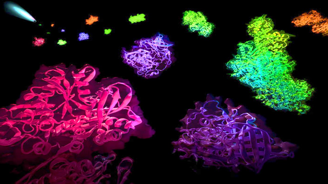
The Triangular Conformation: A Folded Landscape of Inhibition
Beneath the veil of cellular calm, myosin-5a rests in a state of inhibition—a folded, triangular conformation stabilized by an internal architecture both elegant and exacting. In this inactive geometry, the globular tail domain (GTD) folds back to engage its motor heads, forming an intramolecular clasp that suppresses locomotion. Several interactions contribute to this arrested state: electrostatic bonds between the motor domain’s acidic Asp136 and the GTD’s basic Lys1730 and Lys1803, hydrogen bridges between calmodulin and tail, and mechanical coupling via the coiled-coil regions that interlink the two heads and tails. Each contact operates like a latch, reinforcing the motor’s dormancy.
The GTD-GTD interaction, in particular, emerges as a previously underappreciated pillar in this fold. When the N-terminal extensions of each GTD interlock in a head-to-head configuration, they create an inhibitory interface that not only tightens the fold but also masks critical binding regions. This masking effect preserves the protein in a low-energy conformation until activation signals—either ionic or proteinaceous—warrant unfolding.
Experimental constructs that excised specific coiled-coil segments led to the unraveling of this triangulated inhibition. Constructs like His-Myo5a-T1344, retaining segments 1344–1432, preserved dimeric architecture and potent inhibition. In contrast, truncations beyond Lys1432 yielded monomeric tails incapable of stabilizing the same repressive structure. In essence, the GTD dimer acts not merely as a structural consequence of coiled-coils—it is a prerequisite for the suppression of motility.
Intriguingly, mutations at Arg1490 and Lys1491—two conserved residues central to this GTD-GTD clasp—abolish the repression without impairing dimerization itself. This strongly implies that the head-to-head dimer is more than a structural alignment; it is a functionally encrypted interface with profound influence over the entire myosin architecture.
This layered arrangement of folding, stabilized by inter- and intra-domain electrostatics, carves a binary molecular landscape. Myo5a exists in either an inhibited, triangulated rest or an extended, motile activation—each conformation dictated by the interplay between the GTD and its surroundings. The ability to lock this protein in stasis sets the stage for exquisitely timed cellular logistics.
Binding Site Exposed: The Dual State of Inhibited Myosin-5a
A singular elegance emerges when regulatory systems contain built-in redundancy. In the case of myosin-5a, the globular tail domain doesn’t merely inhibit its motor—it also regulates the binding of its activating partner, melanophilin (Mlph). This duality reveals an architecture governed by allostery, where one binding event modulates the availability of another, like a mechanical toggle with opposing states.
The data suggest that Myo5a does not exist in a single inhibited conformation, but rather oscillates between two interconverting inhibitory states: the deeply folded state and a preactivated state. In the former, the GTD-GTD dimer masks the Mlph-binding site, creating a structural occlusion. In the latter, the same binding interface becomes partially exposed, ready to receive an incoming Mlph signal. This oscillation allows for a highly regulated checkpoint before activation.
Upon mutation of Arg1490 and Lys1491—key residues anchoring the GTD-GTD clasp—the frequency of this preactivated state appears to increase. These mutations do not impair the dimeric formation of the GTD per se but rather disturb the interface’s rigidity, rendering the Mlph-binding site more frequently exposed. This state, though still structurally triangulated, becomes chemically permissive to activation.
This allosteric looseness enhances the binding of Mlph-GTBDP, a 26-residue activating peptide, and dramatically lowers the effective Kd. The mutated Myo5a binds Mlph-GTBDP with far greater avidity than the wild-type protein, shifting the activation equilibrium sharply toward motility. The system, once locked, now teeters more readily toward action.
Thus, the folded and preactivated states represent an equilibrium—a molecular switch whose bias can be tuned by structural mutations. In this duality lies the beauty of Myo5a’s control: it is not passively awaiting signals but probabilistically scanning for readiness. This fine-tuned balance sets the foundation for understanding how cargo selection, intracellular location, and signaling converge on a single molecular node.
A Paradox in the Tail: Mutation Weakens Inhibition but Does Not Disrupt Dimerization
The GTD’s ability to dimerize via upstream coiled-coils is critical to the repressive posture of Myo5a. But what happens when key residues at the GTD-GTD interface are removed—not from the structural hinge but from the contact surface itself? The answer reveals a paradox that refines our understanding of protein domain interplay.
R1490A/K1491A mutations, located within the N-terminal extension of the GTD, were designed to disrupt key electrostatic contacts with the opposing GTD. Strikingly, these mutations significantly reduced Myo5a’s ability to repress its motor activity. However, they did not alter the dimeric state of the tail, as confirmed by size exclusion chromatography. This dissociation between dimerization and functional inhibition implies that GTD dimerization is necessary but not sufficient for motor repression—it must also be properly oriented.
Thus, the inhibitory potential of the GTD is governed not merely by proximity or valency but by geometry. Dimerization without correct orientation results in a loss of allosteric leverage. This distinction becomes vital in understanding the impact of small sequence changes on macromolecular behavior—amino acid residues can serve as hinges, locks, or levers depending on their placement in the architectural landscape.
Additionally, when this mutated GTD is placed within full-length Myo5a, the ATPase activity—normally silenced by tail inhibition—is elevated, particularly under EGTA conditions that simulate low calcium. This implies that the tail, unable to hold the motor domain in place, cannot maintain the folded state.
This decoupling of dimerization and inhibition uncovers a new layer of protein logic: structure alone is not destiny. Function depends on coordinated orientation and conformational dynamics that extend beyond domain interactions into the choreography of the entire protein complex. The tail may be connected—but unless it’s clasped just right, Myo5a escapes its leash.
Activation Through Allostery: Melanophilin as a Molecular Wedge
If inhibition is a clasp, then activation is a wedge. Melanophilin (Mlph), the adaptor that links myosin-5a to its cargo in melanocytes, activates Myo5a by prying apart its internal bonds. It achieves this not through brute force but by exploiting structural weakness—binding allosterically at a site where the GTD interacts with the motor domain.
This mechanism centers on Mlph-GTBDP, a short peptide from the GTD-binding domain of Mlph. Though only 26 amino acids long, it binds with precision to a surface that overlaps the GTD-GTD dimer interface. This strategic positioning allows Mlph-GTBDP to allosterically destabilize the GTD’s grip on the motor heads. Once bound, the GTD can no longer enforce its repression.
In mutated Myo5a-FL containing R1490A/K1491A, the binding site becomes more exposed, and the dissociation constant for Mlph-GTBDP drops substantially. The full-length motor now enters an extended, active conformation with greater ease. This suggests that dimeric GTDs in their wild-type state are preconfigured to resist such displacement—until, of course, the system toggles into a permissive conformation.
Notably, this allosteric activation does not occur in the monomeric tail construct, His-Myo5a-T1344, even with the R1490A/K1491A mutation. Here, the absence of additional inhibitory interactions from the motor domain means the GTD is already loosely configured, rendering the peptide’s effect minimal. The full motor must be folded for Mlph-GTBDP to act as a trigger.
Thus, Mlph does not merely bind Myo5a—it pries open a folded machine by allosteric incursion. It is a molecular wedge, targeting not the motor but its repressor, and in doing so, reveals the multi-tiered logic of activation. This insight reframes our understanding of motor protein regulation: control is not linear, but architectural, with signals navigating through a landscape of occlusions and hinges.
The Residue Code: Arg1490 and Lys1491 as Structural Levers
At the core of Myo5a’s regulatory sophistication lies a duet of conserved residues—Arg1490 and Lys1491—embedded within the N-terminal extension of the globular tail domain (GTD). Their precise hydrogen bonding network echoes an allosteric communication channel, enabling or disabling interactions with the myosin heads depending on their conformational state. These residues occupy spatial positions that mimic the hydrogen-bonding interactions formed by melanophilin’s GTD-binding peptide (Mlph-GTBDP), offering a striking molecular mimicry within the protein’s own architecture. This internal mimicry may serve as a protective feedback mechanism, preventing premature activation in the absence of cargo.
Mutagenesis experiments that substituted these basic residues with alanine—resulting in R1490A and K1491A mutants—offered a window into their critical regulatory role. The removal of these positive charges severely weakened the ability of dimerized GTDs to inhibit Myo5a’s ATPase activity. However, these mutations had no comparable effect on monomeric GTDs, confirming that their role is specifically entangled with dimeric-state functionality, possibly stabilizing dimer conformation via electrostatic complementarity. The experimental data implicate these residues as central in maintaining inhibitory tone—when removed, the GTDs lose their grip on the motor domains.
These observations bolster the head-to-head dimerization model. The mutated residues mirror the locations of key Mlph-GTBDP-interacting residues in structural studies. Thus, when dimerized, the GTDs effectively mask their own binding sites for Mlph, locking the protein into its folded, inhibited conformation. This self-repression via structural occlusion is disrupted upon mutation, which functionally “opens” the protein to external regulation by cargo.
Furthermore, pulldown assays reveal that R1490A/K1491A mutations enhance the binding of Mlph-GTBDP to full-length Myo5a, lending credence to a competitive displacement model. The mutant form tilts the equilibrium toward the preactivated state, suggesting a destabilized dimer interface that enables easier access to allosteric sites by cargo proteins. While this mutation increases Myo5a’s responsiveness to Mlph in the full-length protein, it surprisingly does not change the interaction strength in isolated tail constructs, pointing again to the unique structural context of full-length folding.
What emerges is a nuanced view: Arg1490 and Lys1491 do not act in isolation. They are mechanistic levers in a larger, tensioned framework of allosteric inhibition, modular protein folding, and cargo-induced liberation. In their absence, the regulatory architecture loosens, offering a more permissive, activated phenotype.
From Inhibition to Ignition: Mlph-GTBDP and Allosteric Activation
Melanophilin (Mlph), a critical cargo adaptor in melanocytes, binds Myo5a through two separate domains—EFBD and GTBD. Within the GTBD lies a minimal 26-amino acid region, Mlph-GTBDP, which alone can activate Myo5a’s motor function. This activation is not merely a case of steric unblocking but a genuine allosteric mechanism that interrupts GTD’s grip on the motor head, effectively “igniting” the motor from an off state. The spatial separation between Mlph’s GTBDP interaction site and the GTD-motor head interaction site is key here, underscoring a mechanochemical signaling process rather than passive competition.
In full-length Myo5a, R1490A/K1491A mutations significantly enhanced the motor’s responsiveness to Mlph-GTBDP. The ATPase activity of the mutant increased in calcium-depleted (EGTA) conditions—normally associated with the inhibited state—suggesting that the basal repression was weakened. Furthermore, binding assays showed a fourfold increase in Myo5a–Mlph complex formation in the mutant compared to wild-type, implicating Arg1490 and Lys1491 as gatekeepers of ligand accessibility at the GTD dimer interface.
This enhanced binding only occurred in the full-length protein. In isolated tail constructs, particularly His-Myo5a-T1344, the R1490A/K1491A mutation did not affect Mlph-GTBDP interaction, indicating that full-length folding and head-tail contacts stabilize the inhibitory interface. The head domains may exert an additional mechanical constraint, further locking the GTDs into their closed conformation—a conformation that must be destabilized or partially unfolded before cargo binding can proceed.
Mechanistically, this supports a two-state model of activation. In the “folded” state, Myo5a adopts a triangular conformation where GTD-GTD interaction prevents Mlph engagement. In the “preactivated” state, thermal motion or partial conformational loosening exposes the Mlph-binding interface without complete unfolding. Mlph-GTBDP binding then allosterically liberates the motor domains, initiating ATPase turnover and downstream cargo transport.
This transition is exquisitely sensitive to salt concentration. High salt weakens intramolecular ionic interactions, lowering the activation threshold for Myo5a by cargo. Thus, physiological conditions such as ionic microenvironments and local cytoskeletal tension might play hidden roles in regulating motor activation timing in vivo.
Molecular States and Energetic Landscapes of Activation
In capturing the transition from inhibition to activation, the researchers proposed a thermodynamic model involving three states: the folded inhibited conformation (E), a preactivated intermediate (E′), and the fully engaged motor-cargo complex (EL). In this model, the preactivated state is not merely a fleeting conformation but a true thermodynamic basin—one that represents a partially unlatched yet structurally restrained Myo5a ready for ligand engagement.
Mathematically, the steady-state distribution among these three states is governed by four rate constants and their respective dissociation constants. In full-length wild-type Myo5a, multiple internal interactions—head-GTD contacts, coiled-coil backbone stiffness, and GTD-GTD binding—bias the system toward the folded state. However, mutations such as R1490A/K1491A selectively destabilize this folded state, increasing the population of preactivated conformers without wholly unfolding the molecule.
Interestingly, when this model was applied to truncated constructs like His-Myo5a-T1344, the data did not support a significant shift in binding affinity following mutation. In these tail-only contexts, the absence of head-GTD contacts had already destabilized the GTD-GTD interface. As a result, equilibrium already favored the preactivated state, rendering further mutation-induced destabilization inconsequential to cargo binding affinity.
These observations imply that the energetics of Myo5a regulation are context-dependent. In full-length Myo5a, regulation is more tightly controlled, and even slight destabilization of dimer interfaces shifts the molecule toward activation. In contrast, tail constructs are inherently unstable, existing predominantly in states permissive to cargo engagement. These state-specific binding dynamics are central to understanding how Myo5a is selectively activated within the cellular environment.
This model also predicts potential cross-regulation by EFBD, the other Mlph binding region. By binding to exon F in the Myo5a tail, EFBD may itself exert an allosteric pull on the GTD dimer, nudging it toward the preactivated state even before GTBD engages. Thus, dual engagement by EFBD and GTBD might synergize to amplify Myo5a activation, enabling cargo-triggered motility in a spatially restricted and tightly timed fashion.
Toward a Unified Model of Myo5a Regulation
The culmination of these findings provides a structurally coherent and functionally rich model for how Myo5a is regulated at the molecular level. Central to this model is the idea that inhibition and activation are not binary states but conformational equilibria defined by interdomain communication, cargo availability, and local biochemical conditions.
At rest, Myo5a adopts a folded, self-inhibited conformation stabilized by a network of interactions: GTD dimerization via head-to-head contacts, intramolecular hydrogen bonding between GTD and motor domains, and structural constraints enforced by calmodulin-loaded IQ motifs. This inhibition is functionally necessary to conserve ATP and avoid nonspecific actin engagement during motor dormancy.
Upon cargo arrival—especially under permissive ionic conditions—Myo5a transitions through a preactivated state, partially loosening GTD interactions without full dissociation. It is in this poised condition that cargo molecules like Mlph-GTBDP engage, allosterically releasing the GTD’s inhibitory grip. The kinetic and structural changes that follow allow Myo5a to adopt an extended conformation, resume ATPase cycling, and proceed along actin filaments with high processivity.
The role of conserved residues like Arg1490 and Lys1491 is particularly noteworthy because they represent a molecular control valve embedded within the dimer interface. Their strategic position allows them to form transient hydrogen bonds and electrostatic interactions that stabilize the inhibitory head-to-head GTD conformation. These residues act as both anchors and sensors—anchoring the GTDs together to preserve the folded state, and sensing destabilizing forces such as cargo binding or ionic fluctuation. This dual functionality enables them to modulate Myo5a’s state between quiescence and motility with exquisite sensitivity.
Experimental mutagenesis replacing these residues with alanine produced a weakened inhibitory phenotype and a marked enhancement of cargo-induced activation, but only in the context of full-length Myo5a. This conditional effect further underscores the integrative nature of Myo5a regulation, in which the GTDs operate not as isolated units but as modules whose behavior is deeply context-dependent. The architectural integrity of the entire molecule—from motor domain to tail—is required for these residues to exert their full regulatory influence.
In this light, the residues may be viewed as part of a larger “gating motif,” an evolutionarily conserved microdomain within Myo5a that governs allosteric transitions. Their interaction with the Mlph-binding surface creates a steric blockade, and their removal allows greater exposure of this surface, shifting the conformational ensemble toward a state competent for cargo engagement. It is not merely the presence or absence of these residues that controls motor activity, but how their removal alters the energetic landscape of GTD-motor domain interactions and inter-GTD symmetry.
These findings also have significant implications for the understanding of cargo-specific activation mechanisms in other class V myosins. Given the structural conservation across Myo5a, Myo5b, and Myo5c, it is likely that similar regulatory architectures and conformational equilibria are at play in other isoforms. The Arg-Lys motif at the GTD interface may thus represent a universal regulatory switch, fine-tuned by the cellular milieu and post-translational modifications to meet the unique demands of intracellular cargo delivery.
Ultimately, these residues exemplify the delicate balance between structure and function in molecular motors. Their contribution extends beyond simple inhibition or activation; they orchestrate a symphony of conformational states that allow Myo5a to respond to cellular cues with both precision and adaptability. Understanding their role may open avenues for designing targeted interventions in diseases where Myo5a function is impaired, such as in certain forms of albinism, neurodevelopmental disorders, or vesicular trafficking defects.
Study DOI: https://doi.org/10.1074/jbc.M116.724328
Engr. Dex Marco Tiu Guibelondo, B.Sc. Pharm, R.Ph., B.Sc. CpE
Editor-in-Chief, PharmaFEATURES

Subscribe
to get our
LATEST NEWS


Related Posts

Molecular Biology & Biotechnology
The Heart’s Hidden Architects: Reprogramming Cardiac Regeneration Through Intracellular Stem Cell Engineering
Cardiac regeneration, once dismissed as a biological impossibility, has now become a question not of “if,” but of “how.”
Drug Discovery Biology
Unlocking GPCR Mysteries: How Surface Plasmon Resonance Fragment Screening Revolutionizes Drug Discovery for Membrane Proteins
Surface plasmon resonance has emerged as a cornerstone of fragment-based drug discovery, particularly for GPCRs.

Read More Articles

Medicinal Chemistry & Pharmacology
April 15, 2025
Designing Better Sugar Stoppers: Engineering Selective α-Glucosidase Inhibitors via Fragment-Based Dynamic Chemistry
One of the most pressing challenges in anti-diabetic therapy is reducing the unpleasant and often debilitating gastrointestinal side effects that accompany α-amylase inhibition.