Revisiting Nutrient Utilization in Tumor Biology
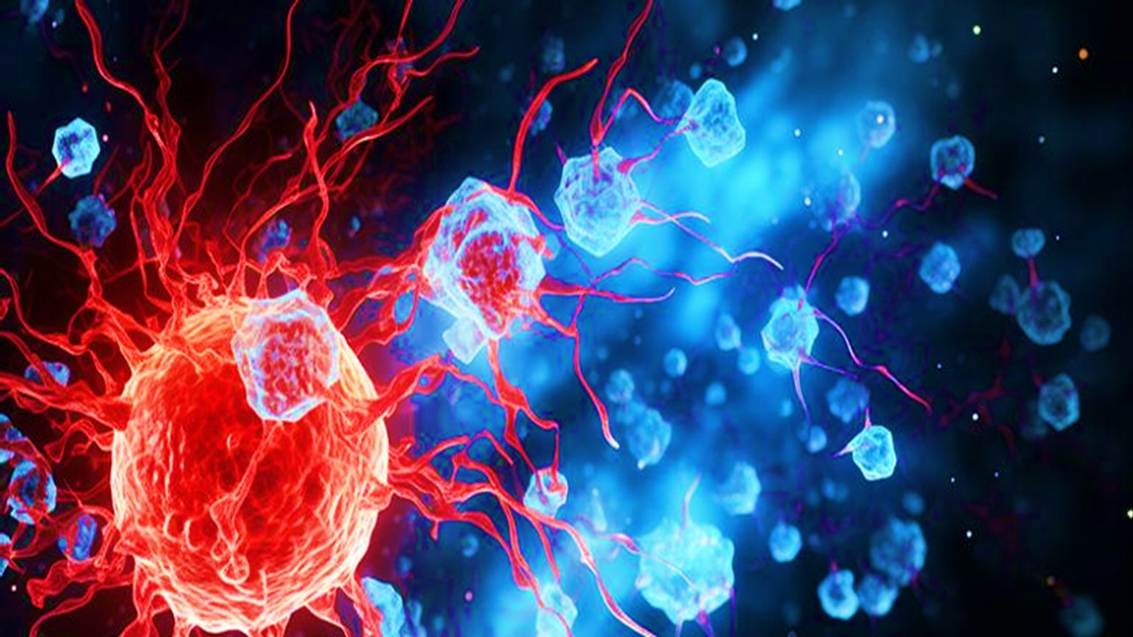
The tumor microenvironment (TME) is a complex, heterogeneous ecosystem where cancer cells and infiltrating immune cells engage in distinct metabolic programs to support their respective functions. Tumor cells preferentially consume glucose via aerobic glycolysis—a phenomenon known as the Warburg effect—while immune cells exhibit diverse metabolic requirements depending on their subtype and activation state. Despite this, the precise mechanisms and patterns of nutrient partitioning within the TME remain incompletely understood.
Recent research provides significant insights into how glucose and glutamine are differentially accessed and utilized by cancer and immune cells in the TME. Using advanced imaging techniques such as fluorodeoxyglucose (FDG) and fluoroglutamine (18F-Gln) positron emission tomography (PET), along with molecular and cellular analyses, these studies reveal that cell-intrinsic programs drive preferential nutrient acquisition, challenging earlier models of metabolic competition.
Glucose Uptake is Predominantly Mediated by Myeloid Cells in the TME
Contrary to the expectation that cancer cells dominate glucose consumption, findings indicate that myeloid cells exhibit the highest glucose uptake on a per-cell basis within the TME. This pattern was consistent across multiple cancer models and validated through a combination of FDG-PET imaging, autoradiography, and flow cytometry-based quantification.
Comparative Analysis of Glucose Uptake
Subcutaneous tumor models, including murine MC38 and CT26 carcinomas, revealed that tumor-infiltrating myeloid cells exhibit higher glucose uptake per cell compared to cancer cells and T cells. Myeloid subsets, including monocytic myeloid-derived suppressor cells (M-MDSCs) and tumor-associated macrophages (TAMs), demonstrated particularly high levels of FDG avidity. Metabolic assays confirmed that these cells maintained elevated extracellular acidification rates (ECAR) and oxygen consumption rates (OCR), indicative of active glycolysis and mitochondrial respiration.
Spatial and Cellular Determinants of Glucose Uptake
Immunohistochemistry and autoradiography showed no significant spatial differences in glucose availability across the TME, indicating that preferential glucose uptake by myeloid cells is driven by intrinsic metabolic programs rather than differential nutrient access. Flow cytometry analysis further validated the purity and viability of isolated myeloid cell populations, confirming that their high glucose uptake is not an artifact of experimental processing.
Implications for Metabolic Interventions
These findings suggest that targeting myeloid cell metabolism may provide a novel avenue for modulating the TME. By altering glycolytic activity in myeloid cells, it may be possible to influence their immunosuppressive functions and enhance anti-tumor immunity.
Cancer Cells Exhibit a Strong Preference for Glutamine Uptake
While myeloid cells dominate glucose consumption, cancer cells exhibit the highest glutamine uptake within the TME. This metabolic specialization supports the anabolic and redox requirements of rapidly proliferating tumor cells.
Mechanisms of Glutamine Utilization
Glutamine uptake in cancer cells is mediated by specific transporters and enzymes, including SLC1A5 (ASCT2) and glutaminase (GLS). Transcriptional profiling revealed elevated expression of glutamine-related genes such as MYCN and ATF4 in cancer cells compared to immune cells. PET imaging using 18F-Gln demonstrated that glutamine avidity is significantly higher in cancer cells than in myeloid or T cell populations across multiple tumor models, including CT26 and MC38 carcinomas.
Glutamine-Glucose Interplay
Inhibition of glutamine uptake using the ASCT2 inhibitor V9302 not only reduced tumor mass but also led to compensatory increases in glucose uptake across all TME cell types. This observation underscores the interdependence of glucose and glutamine metabolism in the TME. Moreover, glutamine restriction altered the immunophenotype of the TME, including increased polarization of TAMs toward an M2-like phenotype.
Therapeutic Implications
The reliance of cancer cells on glutamine metabolism represents a therapeutic vulnerability. Targeting glutamine uptake or metabolism could selectively impair tumor growth while simultaneously reprogramming immune cell metabolism to favor anti-tumor activity.
mTORC1: A Central Regulator of TME Metabolism
Mechanistic target of rapamycin complex 1 (mTORC1) is a critical regulator of nutrient uptake and metabolic activity within the TME. Its activity was found to be elevated in both cancer and myeloid cells, driving their respective preferences for glutamine and glucose metabolism.
mTORC1 and Metabolic Pathways
Rapamycin treatment reduced mTORC1 activity, as evidenced by decreased phosphorylation of ribosomal protein S6 (pS6) in both myeloid and cancer cell populations. This inhibition corresponded with reduced glucose uptake in myeloid cells and decreased glutamine metabolism in cancer cells. Transcriptional analysis revealed that mTORC1 signaling modulates the expression of key metabolic genes, including those involved in glycolysis and amino acid transport.
Effects on Cellular Phenotypes
Rapamycin-mediated mTORC1 inhibition led to decreased cell size and reduced metabolic activity in TAMs and cancer cells. T cells within the TME retained their phenotypic markers but exhibited reduced activation, highlighting the complex interplay between mTORC1 signaling and immune cell function.
Strategic Targeting of mTORC1
Therapeutic inhibition of mTORC1 offers dual benefits: direct metabolic impairment of tumor cells and reprogramming of immune cell activity. By modulating mTORC1-dependent pathways, it may be possible to disrupt the metabolic crosstalk that sustains tumor progression.
Advanced Imaging as a Tool for Metabolic Profiling
The application of PET imaging with radiolabeled glucose and glutamine analogs has provided unprecedented insights into nutrient partitioning within the TME. These techniques allow for real-time, cell-specific quantification of metabolic activity in vivo.
FDG-PET for Glucose Uptake
FDG-PET imaging revealed that myeloid cells account for a substantial fraction of total tumor glucose uptake, challenging traditional assumptions about cancer cell dominance. This finding is particularly relevant for interpreting PET imaging in clinical oncology, as it highlights the contribution of immune cell metabolism to FDG avidity.
18F-Gln PET for Glutamine Metabolism
Glutamine PET imaging confirmed the selective avidity of cancer cells for glutamine, providing a non-invasive method for assessing tumor metabolic phenotypes. This technique could be instrumental in evaluating the efficacy of glutamine-targeted therapies.
Integration with Therapeutic Strategies
By combining metabolic imaging with targeted therapies, clinicians can monitor treatment responses and adapt therapeutic regimens in real-time. For example, changes in PET signals following mTORC1 inhibition or glutamine transport blockade could serve as biomarkers of therapeutic efficacy.
Future Directions
This research elucidates the cell-intrinsic mechanisms governing nutrient partitioning within the TME, with myeloid cells preferentially consuming glucose and cancer cells relying on glutamine. These findings challenge traditional models of metabolic competition and highlight the role of intrinsic metabolic programming in shaping the TME.
Implications for Cancer Therapy
The differential nutrient preferences of TME cell populations offer novel opportunities for therapeutic intervention. Targeting glutamine metabolism in cancer cells or modulating glycolysis in myeloid cells could disrupt tumor growth and reprogram the TME to enhance anti-tumor immunity.
Centrality of Integrative Research
Further studies are needed to explore the molecular mechanisms linking mTORC1 signaling to nutrient partitioning and to identify additional metabolic vulnerabilities within the TME. The integration of metabolic imaging with functional assays will be crucial for translating these findings into clinical applications.
By advancing our understanding of TME metabolism, this research provides a foundation for developing more precise and effective cancer therapies, tailored to the unique metabolic dependencies of tumor and immune cells.
Study DOI: https://doi.org/10.1038/s41586-021-03442-1
Engr. Dex Marco Tiu Guibelondo, B.Sc. Pharm, R.Ph., B.Sc. CpE
Editor-in-Chief, PharmaFEATURES

Subscribe
to get our
LATEST NEWS
Related Posts

Immunology & Oncology
The Silent Guardian: How GAS1 Shapes the Landscape of Metastatic Melanoma
GAS1’s discovery represents a beacon of hope in the fight against metastatic disease.
Immunology & Oncology
Resistance Mechanisms Unveiled: The Role of Glutathione S-Transferase in Cancer Therapy Failures
Understanding this dual role of GSTs as both protectors and accomplices to malignancies is central to tackling drug resistance.
Read More Articles
Myosin’s Molecular Toggle: How Dimerization of the Globular Tail Domain Controls the Motor Function of Myo5a
Myo5a exists in either an inhibited, triangulated rest or an extended, motile activation, each conformation dictated by the interplay between the GTD and its surroundings.
Designing Better Sugar Stoppers: Engineering Selective α-Glucosidase Inhibitors via Fragment-Based Dynamic Chemistry
One of the most pressing challenges in anti-diabetic therapy is reducing the unpleasant and often debilitating gastrointestinal side effects that accompany α-amylase inhibition.


