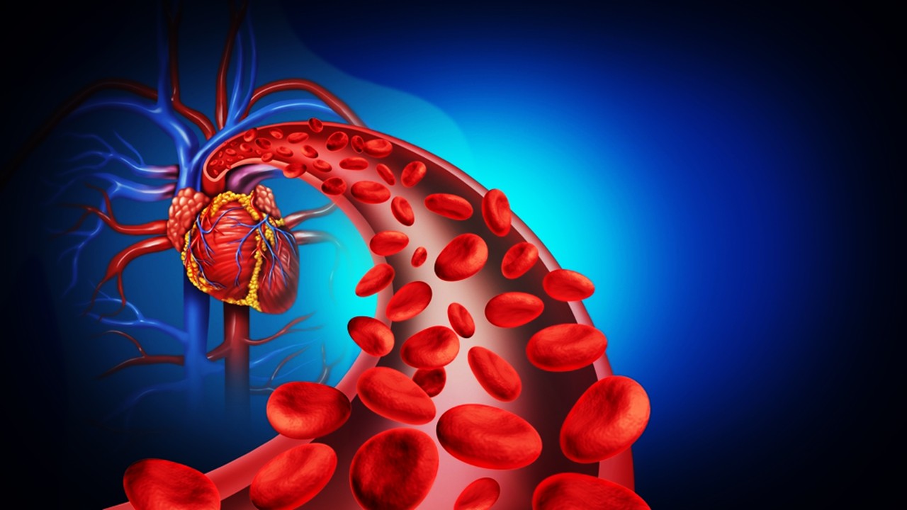
The early choreography: blood films, immune setpoints, and the meaning of osseointegration
The instant a titanium fixture touches living tissue, its fate is negotiated by proteins, not metal, because plasma proteins cloak the surface in a molecular film that sets off coagulation, complement, and platelet programs. That nascent conditioning layer instructs neutrophils and monocytes, which in turn script macrophage polarization dynamics that either perpetuate inflammation or pivot toward constructive remodeling. Within this biochemical theatre, fibrin becomes a provisional matrix that tethers cells, concentrates growth factors, and seeds microgradients that guide osteoprogenitor recruitment. Over hours and days, osteoclasts clear necrotic bone while osteoblast precursors probe the interface, reading adsorbed protein orientation and nanoscale energy cues as if they were signage. The biological prize—bone directly apposed to the implant without intervening fibrous tissue—was formalized decades ago and remains the operational definition of osseointegration. Understanding that this “decision” is made at the surface, and made early, is why modern implant science has shifted from bulk alloys to bioactive interfaces.
The transition from blood clot to woven bone is not a simple cascade but a feedback-controlled process in which cells re-write the very matrix they sense. Platelets and early leukocytes deposit cytokines that alter the conformation of the first-layer proteins, and that conformational rewriting changes integrin engagement thresholds on osteoprogenitors. As these precursors adhere, they deposit collagen-rich matrices whose fibril orientation back-informs their own cytoskeletal alignment and nascent mineral nucleation. Meanwhile, macrophages adjudicate debris clearance and biomaterial recognition, and their phenotype distribution can tilt toward either fibrotic encapsulation or bone formation. The surface free energy and nanotopography of the implant bias that tilt, effectively tuning the setpoint of the local immune response. When the setpoint favors resolution and remodeling, osteoblasts mineralize the interface and lock the fixture in functional continuity with living bone.
Because the interface is both chemical and topographic, protein adsorption kinetics alone cannot predict outcomes; spatial features at the micro–nano scale add an instructive dimension. Adsorbed albumin, fibrinogen, and fibronectin can adopt distinct orientations on hydrophilic versus hydrophobic domains, and these orientations expose or hide integrin-binding motifs. Subtle rearrangements at that layer translate into distinct adhesion-strength profiles for osteogenic cells, changing their mechanotransductive tone. Early mechanosensing modifies nuclear shuttling of transcription factors that govern osteogenic commitment, creating a memory of the surface that persists even as cells move and divide. The result is a self-amplifying loop in which surface design edits the biology that edits the surface-bound matrix. Capturing and steering this loop is the central rationale behind functionalizing implant surfaces rather than merely polishing them.
Historically, osseointegration was discovered as a biological inevitability of certain metals in bone, yet contemporary practice treats it as an engineered outcome. The modern view accepts that immune recognition is not an obstacle but a lever, and that biomaterials can be written to elicit pro-healing immunity. This “osteoimmunomodulatory” perspective recasts surface chemistry, roughness, and adsorbed ligands as tunable variables that steer macrophages, dendritic cells, and osteoprogenitors toward constructive programs. The question is no longer whether titanium integrates, but how predictably, how fast, and under which systemic and local constraints. Answering that question starts with a close read of the bone-healing playbook and continues with designs that speak the biochemical language of that playbook at the interface. The latest syntheses of the field now articulate these design rules with unusual clarity.
Topography as instruction: micro–nano architectures that coach cells
Micro-roughened and nanotextured surfaces behave like passive scaffolds only in appearance; to cells they function as information-rich landscapes. Peaks and valleys at the microscale facilitate fibrin retention and early clot stability, while submicron and nanoscale features gate integrin clustering and focal-adhesion maturation. As focal adhesions mature, osteoprogenitors increase cytoskeletal tension, which feeds forward into osteogenic gene programs and matrix mineral nucleation. In parallel, topographic anisotropy can orient collagen deposition and canaliculi alignment, subtly aligning early tissue architecture with load paths. Because these effects arise from physical cues rather than exogenous factors, they are inherently local and persistent, surviving the washout of soluble signals. This is why micro–nano hybrid surfaces have become the workhorses of contemporary implant design.
Several industrialized routes generate such instruction-rich topographies, each leaving a distinct “accent” on the titanium oxide layer. Sand-blasted and acid-etched surfaces create interlocking microcavities that enhance mechanical interdigitation and early cell adhesion, while maintaining a chemistry hospitable to protein adsorption. Controlled anodization grows ordered oxide nanotube arrays that raise specific surface area and present regular nanoscale cues to integrins and filopodia. Plasma-sprayed textures yield macro-to-micro asperities that can be layered with later chemistries, combining geometric retention with functional coatings. These manufacturing choices are not interchangeable aesthetics; they set the initial conditions that biology elaborates upon. Long-term performance analyses increasingly contextualize outcomes by the micro–nano grammar written at day zero.
As nanotubes, pits, and ridges modulate osteoblast behavior, they also tune immune tone at the interface. Certain nanotube diameters foster macrophage phenotypes associated with wound resolution and matrix deposition, thereby aligning early immunity with osteogenesis. Roughness-driven increases in surface free energy can also amplify spontaneous wetting, which stabilizes the protein layer and improves cell capture. Texture thus serves a dual role: it scaffolds bone mechanics and curates immunity, closing the loop between topography and host response. This is not a static state; as cells colonize and secrete matrix, the interface evolves, so the most effective textures anticipate and support that evolution. The engineering problem is to select topographies that remain instructive as the biological veneer thickens.
Because additively manufactured frameworks can embed multiscale porosity, topology can now be co-designed from the body of the implant out to the oxide skin. Such architectures promote vascular ingress and osteon interlock while simultaneously presenting nanoscale cues at the immediate contact plane. In practice, this means a single implant can couple mechanical compliance gradients with integrin-scale patterning, reducing stress shielding while accelerating biologic fixation. The design space is large, but converges on a principle: write the interface for the first week, and the tissue will write the rest. Even modest shifts in those early instructions can compound across remodeling cycles. Biomimetic topography is therefore best viewed as a language, not a texture.
Inorganic biomimesis: calcium-phosphate chemistries that speak “bone”
Bone’s mineral phase is a carbonate-rich apatite, so coatings that approximate its chemistry can nudge cells toward a familiar conversation. Hydroxyapatite and related calcium-phosphate phases serve as ion reservoirs and nucleation templates, encouraging osteoblasts to mineralize along an already sympathetic lattice. When applied as thin films on titanium, these coatings present both chemical likeness and a gentle dissolution behavior that supplies calcium and phosphate during early healing. The resulting microenvironment stabilizes alkaline phosphatase activity and supports collagen mineralization, effectively lowering the activation energy for early bone apposition. Because the mineral is “recognized” by cells and proteins, it can accelerate the transition from provisional matrix to woven bone. The interface shifts from a metal–protein dialogue to a bone–bone dialogue with far less translation.
How the mineral arrives on the implant matters, because deposition routes imprint crystallinity, porosity, and adhesion strength. Air-plasma spraying yields thick, crystalline layers with substantial roughness that are clinically proven yet vulnerable to interfacial cohesion issues if poorly tuned. Sol-gel routes can generate homogeneous, thinner films with strong bonding and controllable phase composition, supporting stable ion release without excessive brittleness. Electrophoretic and electrochemical methods enable conformal deposition on complex geometries, though they require careful post-processing to avoid overly inert crystals. More recently, bioinspired strategies harness catechol chemistry to seed apatite at physiological conditions, marrying strong adhesion to the metal with a nanogranular, cell-tuned mineral. Each method yields a distinct mineral microstructure that cells read differently.
Nature rarely writes pure hydroxyapatite; minor ions inscribe biological nuance into the lattice. Inspired by that nuance, dopants such as magnesium, strontium, zinc, or fluoride can be woven into coatings to modulate dissolution, osteogenesis, and interfacial remodeling. Substitutional ions subtly distort the crystal lattice and adjust surface charge, which can bias protein adsorption and cell behavior toward pro-osteogenic states. Some ions also add ancillary function, including antimicrobial tone or enhanced early vascular support, without introducing large-molecule complexity. When dopants are combined judiciously, the coating becomes a multifactorial signal rather than a single-note template. In practice, such biomimetic mineral chemistries often outperform undoped apatite for early bone contact.
The mineral phase is not only a signal; it is also a platform for hierarchical design. Apatite coatings can host nanoscale roughness, carry tethered peptides, or embed nucleation islands that further sculpt cell behavior. They can be layered onto specific microtopographies to harmonize geometric retention with biochemical familiarity. In well-designed stacks, topography captures the clot, mineral chemistry stabilizes osteogenic adhesion, and ion release sustains the early metabolic needs of matrix deposition. This orchestration reduces reliance on exogenous biologics while retaining a path to add them when needed. As the field progresses, mineral coatings are evolving from passive films into programmable, living-interface partners.
Organic cues and matrices: ECM-inspired coatings that direct fate
Where mineral says “bone,” extracellular matrix says “how.” Collagen type I, the dominant organic constituent of bone matrix, offers cells integrin-engaging motifs that tune adhesion strength, cytoskeletal tension, and lineage commitment. When covalently anchored to titanium or to mineral layers, collagen establishes a biochemical grammar compatible with osteoprogenitor spreading and ordered fibrillogenesis. The alignment and density of collagen on the surface influence downstream fibronectin assembly, which in turn patterns further matrix deposition and mineral nucleation. These events are not cosmetic; they program early tissue architecture at the interface. Stable collagen coatings have repeatedly been associated with improved bone contact in preclinical and clinical contexts.
ECM is more than collagen, and adding select matrix components can refine the conversation. Laminin-derived motifs accelerate early bone contact in challenging environments by enhancing progenitor capture and early adhesion. Hyaluronan and chondroitin sulfate, when layered with collagen, modulate hydration and growth-factor retention, shaping how cells perceive stiffness and available cues. Such glycomatrix elements also influence macrophage behavior, improving the timing of the inflammatory-to-healing transition. Multicomponent ECM films therefore can serve as tunable, living-like skins over otherwise inert metals. The key is achieving stable immobilization without masking the very motifs that cells are meant to sense.
Covalent coupling strategies have become essential because simple adsorption sheds too quickly to guide weeks-long healing. Silanization chemistries, catechol-based primers, and gentle crosslinkers improve retention while preserving bioactivity of sensitive ECM ligands. These tethers also survive surgical handling and early fluid shear, maintaining signal fidelity during the noisy postoperative period. When ECM cues remain present throughout clot resolution and early osteoid formation, they exert outsized influence on matrix organization and mineral alignment. The outcome is not just faster integration but better organized bone at the contact zone. Durable signaling is what distinguishes decoration from design.
Because surfaces evolve under biologic use, ECM-inspired coatings work best when they anticipate and welcome remodeling. Collagen can be presented in fibrillar or cluster forms, each seeding different modes of fibronectin assembly and stress fiber patterning. As cells lay down their own ECM, the original cues cede control to a self-reinforcing matrix that inherits the initial alignment and spacing. This handoff marks success: the implant’s synthetic language gives way to the host’s. Designing for that handoff—strong enough to start, modest enough to let biology finish—is a practical art at the core of ECM functionalization. Contemporary reviews now codify these principles into actionable surface recipes.
Biofunction on demand: growth-factor and immunoactive interfaces for resilient integration
When topography and base chemistry need help, implants can deliver instructive molecules with temporal control. Bone morphogenetic protein on a mineralized or heparinized carrier can be held at the interface and released gradually, avoiding the burst losses that accompany weak adsorption. Tethering strategies that nano-anchor growth factors or their binding domains directly to a pre-coated surface maintain local concentration during the vulnerable early period. Vascular endothelial cues can be co-presented to support microvessel ingrowth that keeps nascent bone viable as it matures. The common thread is kinetics: the interface must whisper for days, not shout for minutes. With careful pairing of carriers and ligands, that whisper becomes a sustained conversation.
Not every indication benefits from the same biologic, and that is an argument for modularity rather than maximalism. In irradiated or otherwise compromised bone beds, combined osteogenic and angiogenic cues can restore integration trajectories that would otherwise stall. In healthy beds that already respond briskly to topography and mineral chemistry, additional factors may add little and can even distract biology if released too fast. This reality has shifted focus toward covalent immobilization, sequestration via smart glycomatrices, and mineral-incorporated payloads that meter presentation. The aim is not to flood the interface but to time-gate it. Precision in delivery can be as powerful as potency in molecule choice.
As growth-factor engineering matures, immunoactive design has emerged as an equally potent lever. Surfaces that bias macrophages toward pro-healing phenotypes create a permissive niche for osteogenesis without exogenous morphogens. Nanotextures, specific mineral chemistries, and ECM glycans all contribute to this immunologic curation, often with greater robustness than single recombinant factors. The payoff is resilience: interfaces that integrate in smokers, diabetics, or after infection are those that tame inflammation while sustaining progenitor function. By viewing osseointegration as an immune-guided tissue-construction project, designers have opened a wider solution space. The best surfaces are now both osteoinductive and osteoimmunomodulatory.
The field’s trajectory points toward surfaces that are programmable, layered, and diagnostically visible. Additive manufacturing provides porosity and mechanical grading, while oxide nanotubes and apatite films offer nanoscale canvases for biomolecules. Catechol primers and glycomatrix tethers secure cues across the noisy postoperative interval, and modular payloads allow indication-specific tuning without redesigning the base implant. This stack can be monitored and iterated by correlating early biologic readouts with long-term stability, closing the loop between design and outcome. In that future, osseointegration is not left to chance; it is orchestrated across scales and across time. The recent comprehensive syntheses of implant surface science provide a map for building that future now.
Study DOI: https://doi.org/10.3389/fbioe.2023.1320307
Engr. Dex Marco Tiu Guibelondo, B.Sc. Pharm, R.Ph., B.Sc. CpE
Editor-in-Chief, PharmaFEATURES

Subscribe
to get our
LATEST NEWS
Related Posts
Regenerative Medicine & Biomaterials
Programmable Nitric Grafts: Steady Nitric Oxide Orchestrates Small-Vessel Healing
Embedding nitrate-driven nitric-oxide release within PCL grafts programmatically guides vascular regeneration across the lumen, media, and progenitor niche.

Regenerative Medicine & Biomaterials
Bioactive Networks: Photocrosslinkable Gelatin Hydrogels and Cytokine Modulation in Human Immunity
GelMA hydrogels demonstrate that the architecture of matter itself can modulate the architecture of inflammation.
Regenerative Medicine & Biomaterials
Peptide Scaffolds: Programming Biomaterials for Regeneration
Peptide biomaterials are programmable building blocks that rewire scaffolds into active agents of tissue repair.
Regenerative Medicine & Biomaterials
Carbon Conductors, Living Sensors: Graphene Substrates Steer Inner Ear Lgr5+ Progenitors Toward Hair-Cell Identity
Electroconductive graphene scaffolds guide Lgr5⁺ progenitors toward bona fide hair-cell phenotypes by aligning membrane biophysics with lineage-specifying transcriptional cues.
Read More Articles


Medicinal Chemistry & Pharmacology
March 12, 2026
Leaner Molecules: Structural Simplification Outperforming Brute-Force Potency Potency Chasing in Lead Optimization
Structural simplification is the science of turning chemically overbuilt leads into more efficient, drug-like molecules without surrendering their therapeutic logic.
Artificial Intelligence and Data Analytics
March 11, 2026
Digital Stewardship: Governing Access, Transparency, and Accountability in Clinical Data Warehouses
Clinical data warehouse governance determines how integrated health data can be responsibly accessed, shared, and reused to enable modern biomedical research.
Medicinal Chemistry & Pharmacology
March 10, 2026
Pocket Alchemy: Automated Structure-Based Design Rewrites Molecular Invention Logic
Structure-based de novo drug design is the science of teaching algorithms to imagine molecules under the hard constraints of protein geometry and real-world chemistry.
Immunology & Oncology
March 9, 2026
Oncology Rewired: Molecularly Precise Therapeutics Redefining the Architecture of Cancer Treatment
Emerging cancer therapies are redefining treatment by targeting the molecular circuitry, immune interactions, and metabolic vulnerabilities that allow tumors to survive and evolve.
Medicinal Chemistry & Pharmacology
March 6, 2026
Algorithmic Therapeutics: Artificial Intelligence Reshaping Biotech Drug Discovery and Product Development
Artificial intelligence is transforming biotech by making therapeutic discovery less like screening for luck and more like engineering across molecules, biologics, and delivery systems.

Chemistry, Manufacturing & Controls
March 4, 2026
Factory Logic: Modular Pharmaceutical Manufacturing Enabling Rapid Drug Production and Continuous Innovation
Modular pharmaceutical factories transform drug manufacturing into a continuously evolving technological system capable of integrating new therapies, processes, and production innovations without disrupting regulated operations.