Unearthing the First Cellular Movers
In 1985, the scientific community was shaken by a discovery that transformed our understanding of intracellular logistics—kinesins, the first microtubule-based molecular motors. Initially identified in cytoplasm extracted from the squid’s giant axon, these motors defied previous assumptions that intracellular cargo relied solely on diffusion. The isolation of the kinesin-1 complex—a fast, anterograde transport motor—was a landmark event, revealing an elegant molecular machine composed of two identical Kinesin Heavy Chains (KHC) and two distinct Kinesin Light Chains (KLC). This heterotetramer was the first glimpse into the machinery that enables cargo transport along microtubules within cells.
Subsequent discoveries expanded this family of motors beyond kinesin-1, with kinesin-2 emerging from echinoderm embryos. Comprising two non-identical heavy chains and an accessory kinesin-associated protein (KAP), kinesin-2 proved essential for intraflagellar transport during the assembly of cilia—antenna-like organelles crucial for cell signaling. These revelations illuminated the vast cellular network powered by kinesins, leading to the recognition that mammals encode more than 40 kinesin variants, spanning 14 distinct families.
The molecular motors had not only arrived but established themselves as the lifeline for eukaryotic cells, orchestrating a symphony of movements essential for growth, division, and homeostasis.
A Molecular Ballet: Kinesin Structure and Regulation
The diversity within the kinesin family is mirrored in their complex yet elegant structure. At the heart of each kinesin-1 molecule lies a motor domain, or “head,” which is responsible for binding to microtubules and hydrolyzing ATP. The motor domain is followed by a flexible neck linker, which connects to the stalk—a coiled-coil alpha-helical structure that facilitates dimerization by intertwining two heavy chains. At the end of this molecular stalk lies the tail domain, which interacts with the light chains, acting as a tether for cargo.
Kinesin motors achieve motion through a series of conformational changes driven by ATP binding and hydrolysis. The motor domain’s Switch I and Switch II regions are integral to the transmission of mechanical force, acting like levers to synchronize the motor’s attachment to the microtubule with energy release. However, basal activity in kinesins remains low until they engage microtubules. Many kinesins self-regulate through autoinhibition, where the tail domain folds back onto the motor head, preventing premature activity. This inhibition is lifted upon interaction with cargo or adapter proteins, ensuring that energy is conserved until the motor is ready to transport cellular cargo.
Despite their structural similarities to G proteins—enzymes that hydrolyze GTP—kinesins have evolved specialized elements for ATP hydrolysis, reflecting their highly specific roles in intracellular transport. These motors embody an exquisite balance of precision and versatility, adapting to the needs of various cellular environments.
Delivering Life’s Cargo: The Dynamics of Transport
Cells are bustling environments where large cargo—vesicles, organelles, and protein complexes—must be precisely transported to specific destinations. While small molecules such as glucose can diffuse through the cytosol, bulkier cellular components rely on motor proteins like kinesins to navigate the crowded intracellular landscape. Kinesins achieve this by “walking” unidirectionally along microtubule tracks, each step powered by the hydrolysis of one ATP molecule.
Emerging research has challenged the once-held belief that ATP hydrolysis alone propels the kinesin forward. Instead, recent models suggest that the binding of the motor head to the microtubule generates the force necessary for movement, with the trailing head diffusing forward before reattaching. This ingenious coordination ensures that kinesins can carry cellular freight over long distances efficiently and without derailment. Remarkably, pathogens such as the HIV virus exploit this system, hitching rides on kinesins to shuttle viral components within host cells, underscoring the importance of these motors in both health and disease.
The concept of “motor redundancy” further enriches our understanding of intracellular transport. Cells deploy multiple kinesins to move critical cargo, ensuring that if one motor falters, others can compensate. This redundancy guarantees that essential organelles, such as mitochondria, reach their destinations without delay, safeguarding cellular metabolism and signaling.
The Mechanism of Motion: A Molecular Step Dance
In 2023, researchers achieved the first direct visualization of kinesins in action, revealing a fascinating “hand-over-hand” mechanism. Each kinesin molecule consists of two motor heads that alternate positions with every step along the microtubule. The process begins with the trailing head releasing inorganic phosphate, causing it to detach from the microtubule. As the leading head binds ATP, the neck linker docks, propelling the trailing head forward in a sweeping motion that positions it ahead of the leading head.
This intricate dance continues in cycles, with each step precisely synchronized by ATP binding and hydrolysis. The mechanism ensures that kinesins maintain a steady pace, efficiently delivering cargo to its destination. The real-time visualization of these motors offers a glimpse into the molecular choreography that underpins cellular life, transforming our understanding of motor protein function.
Kinesins in Cell Division: Guardians of Chromosomal Order
Kinesins are not only transporters but also crucial regulators of mitosis, the process by which cells divide. During cell division, molecular motors orchestrate the alignment and separation of chromosomes to ensure accurate genetic distribution. Kinesin-5 proteins, for instance, play a pivotal role in mitotic spindle formation by sliding microtubules apart, thereby maintaining spindle length and integrity.
The Kinesin-13 family, on the other hand, specializes in depolymerizing microtubules at their minus ends, facilitating the dynamic rearrangement of the spindle during anaphase. These motors ensure that sister chromatids are pulled to opposite ends of the cell, preventing chromosomal abnormalities. Any disruption in kinesin activity can lead to catastrophic mitotic errors, highlighting the importance of these motors in maintaining genomic stability.
Current Pharmacological Explorations and Pathophysiological Implications
Kinesins have emerged as promising targets for pharmacological intervention, particularly in cancer therapy. Given their pivotal role in mitosis, inhibitors of Kinesin-5—such as ispinesib—are under investigation for their potential to disrupt cell division in rapidly proliferating tumors. By halting the formation of the mitotic spindle, these inhibitors trigger cell cycle arrest, leading to apoptosis in cancer cells.
In addition to cancer, kinesin dysfunction is implicated in neurodegenerative diseases. Mutations affecting kinesin-1 and related motors are associated with hereditary spastic paraplegia, a disorder characterized by progressive weakness and stiffness in the lower limbs. Pharmacological strategies aimed at enhancing kinesin function could offer therapeutic benefits by restoring intracellular transport in affected neurons.
However, the development of kinesin-targeting drugs remains challenging due to the motors’ intricate regulatory mechanisms and their ubiquitous presence in essential cellular processes. Selectivity is key—therapeutics must inhibit disease-related kinesin activity without impairing normal cellular function. Advances in structural biology and biophysics are paving the way for more precise interventions, offering hope for new treatments in oncology and neurology.
The Future of Lead Discovery: Navigating Uncharted Territories
Despite decades of research, many aspects of kinesin regulation and function remain unresolved. New technologies, such as cryo-electron microscopy and single-molecule tracking, are enabling scientists to explore uncharted territories in kinesin biology. These tools offer unprecedented insights into the structural dynamics of motor proteins, revealing the subtle conformational changes that drive movement and cargo recognition.
The road ahead promises exciting developments in both fundamental biology and drug discovery. As researchers continue to decode the molecular language of kinesins, novel therapeutic avenues are likely to emerge, transforming the way we approach diseases rooted in cellular transport defects. From the cytoplasmic highways of neurons to the dividing cells of tumors, kinesins remain at the forefront of modern biology—molecular maestros orchestrating the symphony of life.
Study DOI: https://doi.org/10.1021/acscentsci.4c00937
Engr. Dex Marco Tiu Guibelondo, B.Sc. Pharm, R.Ph., B.Sc. CpE
Editor-in-Chief, PharmaFEATURES

Subscribe
to get our
LATEST NEWS
Related Posts
Molecular Biology & Biotechnology
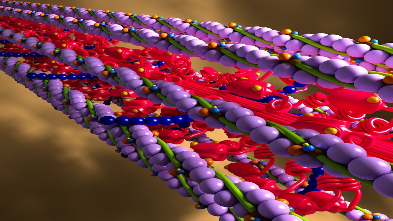
Myosin’s Molecular Toggle: How Dimerization of the Globular Tail Domain Controls the Motor Function of Myo5a
Myo5a exists in either an inhibited, triangulated rest or an extended, motile activation, each conformation dictated by the interplay between the GTD and its surroundings.
Drug Discovery Biology
Unlocking GPCR Mysteries: How Surface Plasmon Resonance Fragment Screening Revolutionizes Drug Discovery for Membrane Proteins
Surface plasmon resonance has emerged as a cornerstone of fragment-based drug discovery, particularly for GPCRs.
Read More Articles
Designing Better Sugar Stoppers: Engineering Selective α-Glucosidase Inhibitors via Fragment-Based Dynamic Chemistry
One of the most pressing challenges in anti-diabetic therapy is reducing the unpleasant and often debilitating gastrointestinal side effects that accompany α-amylase inhibition.


