In the evolving landscape of drug discovery, traditional methods of evaluating efficacy and safety using animal models are being increasingly complemented—and in some cases supplanted—by advanced preclinical models. Emerging technologies now enable the recreation of complex human tissues in vitro, which offers an unprecedented opportunity to study drug interactions in a biologically relevant environment. However, as we embrace these innovative tools, there are several critical design considerations to optimize their integration into the preclinical pipeline.
These considerations range from the source of biological materials used, the complexity captured in 3D culture models, and the challenge of balancing throughput and feasibility. Each of these facets plays a pivotal role in shaping the next generation of lead discovery and advancing therapeutic development.
The Choice of Cellular Materials: The Foundation of Preclinical Models
The choice of cellular material serves as a fundamental cornerstone when designing a preclinical model. Human cell-based systems, often derived from immortalized cell lines, primary human cells, or stem cell-derived sources, form the basis of these models. This decision has profound implications for the relevance, scalability, and application of the model.
Immortalized human cell lines, a staple in drug discovery, provide an enduring, easy-to-manipulate source of cells. Their capacity for indefinite division renders them particularly attractive in terms of consistency and reliability in generating reproducible results. These lines also benefit from a robust background of pre-existing data, which aids in comparative studies. However, their use is not without caveats. The genetic homogeneity that often results from immortalization can obscure the heterogeneity observed in human populations. Moreover, the process of immortalization introduces epigenetic and phenotypic alterations that may limit their biological relevance, especially in models seeking to replicate more nuanced or patient-specific disease mechanisms.
Primary cells, derived directly from human tissues, offer a more biologically relevant alternative but come with their own set of challenges. Variability between patients, limited proliferative capacity, and a rapid loss of phenotype in vitro restrict their widespread use. For diseases with specific genetic underpinnings or toxicity assessments where tissue-specific responses are critical, primary cells may provide an indispensable resource. Yet, the difficulty in scaling and standardizing these cells presents significant obstacles for industrial application.
The emergence of human pluripotent stem cells (hPSCs), including embryonic and induced pluripotent stem cells (iPSCs), provides a novel, standardized, and scalable solution to many of these problems. Derived from adult somatic cells, iPSCs offer researchers a versatile platform capable of generating a wide range of human cell types. This allows for greater flexibility in studying cell-autonomous diseases and testing drug efficacy in a more relevant biological context. However, their immaturity in vitro—often displaying a fetal-like phenotype—presents a critical limitation in modeling adult-onset diseases.
Three-Dimensionality: Adding Depth to Preclinical Models
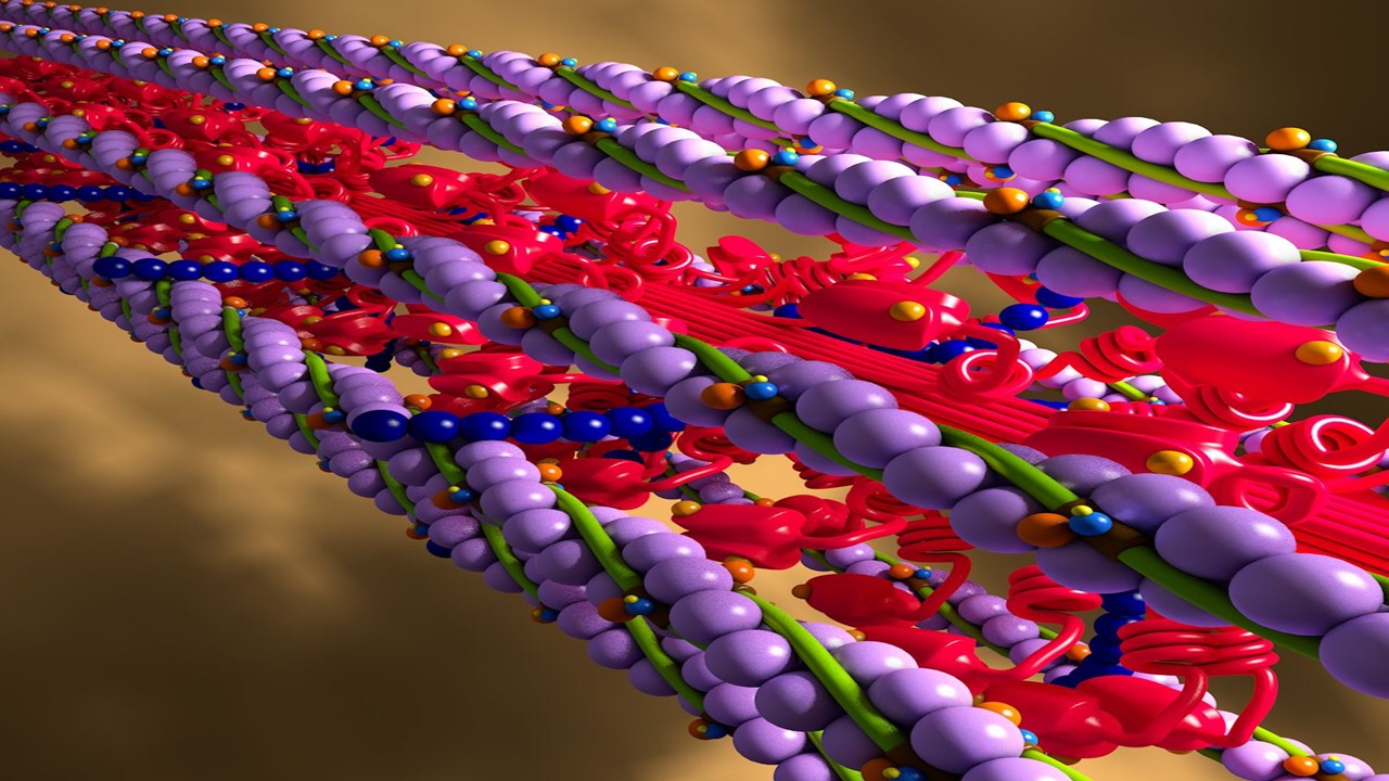
Native tissues do not exist in isolation; they function within highly complex, three-dimensional environments. To recreate these conditions in vitro, researchers have turned to 3D culture models, which more accurately reflect the interactions between cells and their surrounding extracellular matrix (ECM). These models not only facilitate cell-cell communication but also allow for the study of gradient-dependent processes such as nutrient diffusion, mechanical signaling, and drug transport.
Transitioning from 2D monolayers to 3D systems introduces significant complexity into the design and interpretation of preclinical models. Various strategies, such as scaffold-free cell aggregation into spheroids or scaffold-based approaches using ECM hydrogels, have been developed to introduce three-dimensionality into these cultures. Scaffold-free models, which rely on the intrinsic ability of cells to aggregate, are useful for studying diseases where cell-cell interactions and density are critical, such as in cancer or fibrosis. On the other hand, scaffold-based systems that mimic the mechanical and biochemical properties of native ECM are more suitable for studying conditions influenced by cell-ECM interactions, such as tissue stiffness and fibrosis.
However, the introduction of three-dimensionality is not without its own set of challenges. Factors such as porosity, cell density, and the degradation and remodeling capacity of the ECM must be carefully considered. These parameters can fundamentally influence cellular behavior in ways that are difficult to predict from 2D studies. For instance, while substrate stiffness in 2D culture has long been known to regulate cell phenotype and fate, studies in 3D cultures have demonstrated that ECM stiffness alone is insufficient to dictate cellular responses without considering the cell’s ability to deform its surrounding matrix.
The selection of the appropriate ECM material also has profound implications. Natural ECM scaffolds offer biological compatibility but suffer from variability and poor tunability, while synthetic ECMs provide precise control over material properties but often require complex chemical modifications to enhance biological functionality. These materials must not only support cell viability but also allow for the proper morphogenesis of tissues to achieve the level of structural and functional complexity necessary for preclinical drug studies.
Morphogenesis in Tissue Engineering: Building Complex Tissues from the Ground Up
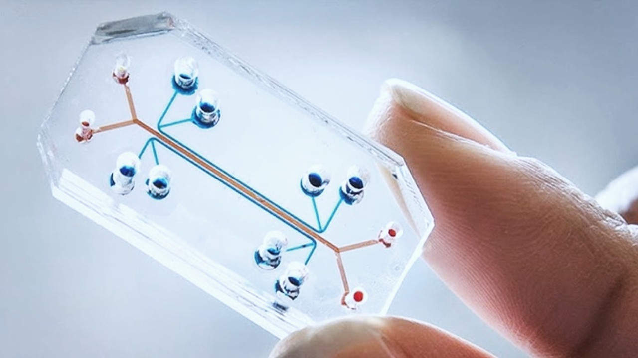
Tissue engineering has been increasingly integrated into the drug discovery process, providing a powerful tool for creating complex biological structures that can mimic human tissues in vitro. To successfully implement tissue engineering in preclinical models, researchers must address a series of critical design considerations. These include selecting the appropriate cell types, designing culture devices that support cellular communication, and validating the model against native tissues.
At the heart of tissue engineering is the process of morphogenesis—the development of organized structures from seemingly random cellular arrangements. To recreate these tissues in vitro, researchers must carefully consider how cells are spatially configured and how their interactions are mediated by the surrounding environment. In many cases, the co-culture of different cell types, including functional cells, stromal cells, and immune cells, is necessary to accurately replicate the in vivo communication networks that sustain tissue function. However, simply placing these cells together is not enough; the culture conditions must be permissive to allow for the natural emergence of tissue-like structures.
Engineered tissues must also be evaluated to ensure that they faithfully recapitulate the functional characteristics of their in vivo counterparts. The use of advanced characterization techniques, such as single-cell RNA sequencing and immunohistochemistry, can provide valuable insights into the degree of tissue morphogenesis and maturity. However, researchers must remain cautious, as single-cell analyses have demonstrated that even the process of isolating primary cells for these assays can significantly alter their transcriptomic profiles.
Despite these challenges, the incorporation of tissue engineering into preclinical drug discovery offers a powerful platform for studying disease mechanisms and evaluating therapeutic candidates in a biologically relevant context.
Space-and Time-Variable Properties of Dynamic Cultures
Human tissues are inherently dynamic, experiencing and responding to a range of mechanical and chemical signals that vary both spatially and temporally. These signals include gradients and fluctuations in metabolites, nutrients, growth factors, and cytokines, as well as mechanical forces from cellular activities and tissue growth. Traditional static in vitro systems often fail to capture these dynamic aspects, potentially limiting their predictive value.
Dynamic Chemical Signals
In vivo, tissues are exposed to dynamic changes in chemical cues, such as the gradients and concentrations of metabolites and growth factors. For instance, tissue repair and regeneration involve fluctuating levels of these factors, which influence cellular responses and drug interactions. Static cultures, on the other hand, do not mimic these fluctuations, leading to potential discrepancies between in vitro and in vivo results.
To address this, preclinical models should incorporate dynamic features that emulate these chemical signals. This could involve systems that allow for controlled variations in the concentration of growth factors and other critical molecules over time, providing a more accurate representation of the in vivo environment.
Mechanical Signals and Tissue Morphogenesis
Mechanical forces, such as those from cellular contraction, migration, and tissue growth, play a crucial role in tissue function and development. These forces contribute to tissue morphogenesis and can influence how cells respond to drugs. Static cultures do not capture these dynamic mechanical signals, which can limit their ability to model complex tissue behaviors.
Incorporating mechanical cues into in vitro models, such as through the use of bioreactors that apply mechanical strain or shear stress, can enhance the physiological relevance of these systems. This approach helps simulate the forces experienced by tissues in vivo, improving the model’s ability to predict drug responses and tissue behaviors.
Transport Challenges in Large 3D Cultures
As in vitro models increase in size and complexity, managing dynamic transport processes becomes more challenging. Large 3D cultures require effective regulation of oxygen and nutrient delivery, as well as removal of waste products, to avoid necrosis and maintain cell viability. The use of bioreactors and perfusion systems can help address these issues by introducing fluid flow and optimizing nutrient and waste exchange.
Moreover, incorporating transport barriers and interconnecting multiple 3D tissue models can provide a more comprehensive representation of drug distribution and metabolism across different organ systems. This approach can enhance the predictive value of the model by better simulating the dynamics of drug interactions within the human body.
Model Feasibility and Likelihood of Adoption
While the biological relevance of preclinical models is crucial, their practical feasibility and likelihood of adoption in drug development workflows are also significant considerations. Several factors influence the feasibility of a preclinical model:
Availability and Standardization of Materials
The source of cells, culture devices, and ECM materials must be readily available, scalable, and standardizable. Models that rely on complex or poorly standardized materials may face challenges in widespread adoption. For instance, the use of novel synthetic ECMs or customized culture devices can complicate model implementation and limit their utility in broader drug development contexts.
Integration with Existing Workflows
Preclinical models should complement and integrate with existing drug development workflows. This includes considering how new models can enhance or augment current systems rather than replace them entirely. For example, while sophisticated 3D models may provide valuable insights, they should be designed to fit within the established framework of drug testing and validation processes.
Balancing Complexity and Throughput
Increasing the complexity of a preclinical model can enhance its biological relevance but may also reduce throughput and ease of use. Models that are too complex may present practical challenges in terms of assembly, operation, and data collection. Therefore, it is essential to strike a balance between model complexity and practicality. For instance, if endothelial barrier function is critical, a simpler Transwell® assay might suffice, whereas studies focused on angiogenesis might require more complex vasculature models.
Measurement and Output Collection
The ability to accurately measure and collect data from a preclinical model is crucial for its success. Models with high complexity may encounter difficulties in obtaining reliable measurements, such as cell viability in 3D cultures. Incorporating genetically engineered readouts, such as fluorescence or luminescence, can facilitate high-throughput measurements, though this may be more challenging with primary cells compared to immortalized cell lines.
In conclusion, the design of new preclinical models should carefully consider both their biological relevance and practical feasibility. Balancing these factors will be essential in developing models that are not only scientifically robust but also widely adopted and useful in the drug development process.
Engr. Dex Marco Tiu Guibelondo, B.Sc. Pharm, R.Ph., B.Sc. CpE
Editor-in-Chief, PharmaFEATURES

Subscribe
to get our
LATEST NEWS


Related Posts
Molecular Biology & Biotechnology
Myosin’s Molecular Toggle: How Dimerization of the Globular Tail Domain Controls the Motor Function of Myo5a
Myo5a exists in either an inhibited, triangulated rest or an extended, motile activation, each conformation dictated by the interplay between the GTD and its surroundings.
Drug Discovery Biology
Unlocking GPCR Mysteries: How Surface Plasmon Resonance Fragment Screening Revolutionizes Drug Discovery for Membrane Proteins
Surface plasmon resonance has emerged as a cornerstone of fragment-based drug discovery, particularly for GPCRs.


Read More Articles

Medicinal Chemistry & Pharmacology
April 15, 2025
Designing Better Sugar Stoppers: Engineering Selective α-Glucosidase Inhibitors via Fragment-Based Dynamic Chemistry
One of the most pressing challenges in anti-diabetic therapy is reducing the unpleasant and often debilitating gastrointestinal side effects that accompany α-amylase inhibition.