The Rise of Organoids: Mimicking Human Organs in a Dish
Organoids, three-dimensional (3D) cellular constructs, are revolutionizing our understanding of human biology by replicating the cellular complexity and architecture of organs. Over two decades, researchers have made substantial strides in cultivating organoids derived from adult stem cells and tumor samples. These models effectively mimic patient-specific characteristics, including genetic variability, protein expression, and tissue microstructures.
Despite these advancements, replicating the full complexity of human organs remains a challenge. Introducing stromal and vascular components, alongside engineering specific sizes and shapes, is crucial to elevating the fidelity of organoid functionality. For example, tubular intestinal organoids have demonstrated the importance of shape in fostering specialized cells and spatial organization akin to in vivo structures. These breakthroughs hint at a transformative frontier for understanding disease mechanisms and testing therapies.
Advancements in Organoid and Tumoroid Synthesis Techniques
The development of organoids and tumoroids has no doubt revolutionized biomedical research by providing 3D models that closely mimic the architecture and functionality of human tissues and tumors. These models are typically derived from pluripotent stem cells, induced pluripotent stem cells, or adult stem cells, which are cultured in specialized conditions that promote self-organization and differentiation. A critical component in this process is the use of extracellular matrix (ECM) hydrogels, such as Cultrex Basement Membrane Extract (BME), which provide a scaffold that supports cell growth and organization . By embedding cells within these hydrogels and supplying a cocktail of growth factors—including WNT, Noggin, R-spondin, and epidermal growth factor (EGF)—researchers can guide the formation of organoids that replicate the complex structures of specific organs.
In cancer research, tumoroids offer a more accurate representation of tumor biology compared to traditional two-dimensional (2D) cell cultures. These 3D models maintain the genetic and phenotypic characteristics of the original tumors, including cellular heterogeneity and microenvironmental interactions. To enhance the physiological relevance of tumoroids, co-culture systems have been developed that incorporate various cell types present in the tumor microenvironment, such as fibroblasts, immune cells, and endothelial cells. These co-culture models enable the study of complex cell-cell interactions and the evaluation of therapeutic responses in a setting that closely resembles in vivo conditions.
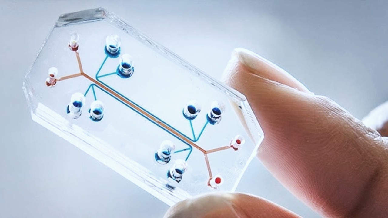
Advancements in bioengineering have further refined organoid and tumoroid synthesis. Techniques such as microfluidic cell culture and bioprinting allow for precise control over the microenvironment, facilitating the creation of more uniform and reproducible 3D structures. For instance, microfluidic devices can simulate physiological conditions by providing controlled nutrient flow and waste removal, thereby supporting the long-term culture of organoids and tumoroids. Additionally, the integration of vascular networks within these models addresses challenges related to nutrient diffusion and waste elimination, enhancing their viability and functionality. These technological innovations are pivotal in advancing the application of organoids and tumoroids in disease modeling, drug discovery, and personalized medicine.
Tumoroids: A Window into Cancer Invasion
Cancer invasion, a hallmark of malignancy, involves tumor cells spreading into surrounding tissues through various migration modes. Whether as individual cells navigating their environment or as cohesive groups propelled by “leader cells,” these dynamics are key to understanding tumor progression. Collective migration, prominent in breast and lung cancers, relies heavily on the mechanical forces exerted by the tumor’s geometry.
To unravel these mechanisms, researchers are increasingly turning to tumoroids—3D cancer spheroids that replicate tumor heterogeneity. However, achieving non-spherical tumoroids has proven elusive, despite the evidence that geometry plays a pivotal role in cancer invasion. Geometric cues in simpler 2D cultures have shown that specific shapes significantly influence cellular behavior, from transcription factor expression to migration patterns. Expanding this understanding to 3D tumoroids could revolutionize cancer biology and therapy.
Introducing the ReSCUE Platform: A Game-Changer in 3D Tumoroid Engineering
The Recoverable-Spheroid-on-a-Chip with Unrestricted External Shape (ReSCUE) platform marks a significant leap in cancer research technology. This microfluidic device facilitates the formation of tumoroids and organoids in various shapes, their targeted release, and downstream analysis. Building on a hydrodynamic droplet-trapping mechanism, the ReSCUE platform introduces a dual-layer design that integrates customizable microwells with fluidic channels.
The process begins with cell suspensions loaded into shape-specific microwells coated with a hydrogel precursor. After brief incubation, the cells form 3D constructs, which can then be gently released for further study. This innovative approach overcomes the limitations of traditional methods by enabling precise control over shape and ensuring structural integrity during culture and recovery.
Tumoroid Formation: Shape, Structure, and Survival
Using the ReSCUE platform, researchers have successfully generated tumoroids in disk, rod, oval, U, and I shapes. Breast cancer cell lines and patient-derived organoids (PDOs) displayed high viability and shape fidelity, confirmed through confocal microscopy and immunostaining. These studies revealed well-defined cytoskeletal structures and cell-cell junctions, critical for understanding tumor mechanics.
The geometry of these tumoroids profoundly influences their biological behavior. For example, rod-shaped tumoroids exhibited enhanced cell proliferation and the formation of multicellular strands at high-curvature regions, mimicking invasive tumor fronts. U-shaped tumoroids displayed similar tendencies, with cells expanding preferentially in positive curvature areas while maintaining structural cohesion.
Long-Term Culture in Unconstrained Environments: Unraveling Shape Dynamics
Tumoroids released from the ReSCUE platform into a biomimetic hydrogel matrix exhibited remarkable growth over 21 days. Disk-shaped tumoroids retained their original geometry, while rod and U shapes developed distinctive multicellular strands from high-curvature regions. These findings highlight the role of geometry in driving collective invasion patterns.
Interestingly, areas with positive curvature fostered higher cell density and proliferation, while negative curvature regions showed reduced activity. These observations align with the mechanical forces exerted during tumor progression, where cells in positive curvature zones propel forward, creating invasive fronts.
Patient-Derived Organoids: Bridging the Gap Between Models and Reality
The ReSCUE platform’s versatility extends to PDOs, derived directly from breast cancer patients. Disk, rod, and U-shaped PDOs were cultured, released, and maintained for 21 days, offering a unique opportunity to study patient-specific tumor dynamics. Similar to cell line-based tumoroids, these PDOs exhibited geometry-driven growth patterns, with high-curvature regions driving proliferation and strand formation.
Immunostaining of PDOs revealed key markers of cellular behavior, including E-cadherin and Ki-67, which correlate with cell junction integrity and proliferation, respectively. These findings reinforce the importance of PDO shape in modeling tumor invasion and provide a framework for personalized cancer research.
Tumor Shape and Clinical Implications
Tumor shape has long been recognized as an indicator of malignancy in clinical imaging. Irregularly shaped tumors often correlate with higher invasiveness and poorer prognosis. The ReSCUE platform’s ability to replicate these shapes in vitro opens new avenues for studying the interplay between tumor geometry and clinical outcomes.
Beyond fundamental research, these models hold potential for refining therapeutic strategies. For instance, understanding how geometric cues influence drug response could lead to more targeted treatments. Moreover, ReSCUE’s capacity to generate uniform, shape-specific tumoroids provides a robust tool for drug screening and radiation therapy optimization.
Toward a New Era in Tumor Modeling
The ReSCUE platform represents a paradigm shift in 3D tumor modeling, offering unprecedented control over shape and enabling long-term studies in unconstrained environments. Its applications extend beyond cancer research, with potential use in organoid tissue engineering, regenerative medicine, and drug discovery.
By bridging the gap between traditional 2D cultures and the complexities of in vivo tumors, ReSCUE provides a powerful tool to unravel the intricacies of cancer biology. As research advances, this platform could pave the way for groundbreaking discoveries in cancer invasion, therapy resistance, and patient-specific treatments. Shape, as it turns out, is more than a visual characteristic—it’s a key driver of life and disease.
Study DOI: https://doi.org/10.1002/adma.202410547
Engr. Dex Marco Tiu Guibelondo, B.Sc. Pharm, R.Ph., B.Sc. CpE
Editor-in-Chief, PharmaFEATURES

Subscribe
to get our
LATEST NEWS
Related Posts

Immunology & Oncology
The Silent Guardian: How GAS1 Shapes the Landscape of Metastatic Melanoma
GAS1’s discovery represents a beacon of hope in the fight against metastatic disease.
Immunology & Oncology
Resistance Mechanisms Unveiled: The Role of Glutathione S-Transferase in Cancer Therapy Failures
Understanding this dual role of GSTs as both protectors and accomplices to malignancies is central to tackling drug resistance.
Read More Articles
Myosin’s Molecular Toggle: How Dimerization of the Globular Tail Domain Controls the Motor Function of Myo5a
Myo5a exists in either an inhibited, triangulated rest or an extended, motile activation, each conformation dictated by the interplay between the GTD and its surroundings.
Designing Better Sugar Stoppers: Engineering Selective α-Glucosidase Inhibitors via Fragment-Based Dynamic Chemistry
One of the most pressing challenges in anti-diabetic therapy is reducing the unpleasant and often debilitating gastrointestinal side effects that accompany α-amylase inhibition.


