MicroRNAs as Biological Signatures
MicroRNAs (miRNAs) are short, non-coding RNA molecules that regulate gene expression post-transcriptionally and have emerged as key biomarkers in oncology. Their dysregulation is strongly linked to tumor initiation, progression, and metastatic behavior, making them powerful signals for early-stage cancer detection. Unlike proteins or metabolites that vary widely between tissues, miRNAs often display highly conserved and reproducible expression patterns in diseased states. However, their abundance in circulation is remarkably low, requiring sophisticated methods for extraction and quantification. Conventional assays such as northern blotting or qRT-PCR have established sensitivity but demand costly equipment, technical expertise, and tightly controlled laboratory conditions. These limitations motivate the development of alternative platforms that couple selectivity with ease of use.
Biosensors offer a compelling solution by directly translating molecular interactions into quantifiable outputs without the constraints of bulky instrumentation. Early iterations of biosensors utilized fluorescence or electrochemical readouts, but these approaches retained dependencies on controlled environments and expensive detectors. In contrast, visual and photothermal detection platforms represent an advance toward point-of-care feasibility. By transforming a hybridization event into a temperature change readable by a standard thermometer, one can simplify detection while maintaining diagnostic precision. This shift embodies the broader trend in biotechnology: decentralizing diagnostic capability without sacrificing analytical rigor. Such innovations align with the demands of modern healthcare systems that prioritize accessibility and rapid response.
The intrinsic challenge with miRNAs lies in their fragility; they are easily degraded by nucleases, which hampers reliable detection. This instability complicates both storage and processing, often forcing researchers to employ stringent RNase-free conditions and chemical stabilizers. Furthermore, sequence similarities between different miRNA families pose selectivity issues, as cross-reactivity undermines confidence in biomarker validation. Biosensor design must therefore integrate structural recognition strategies that discriminate among highly homologous sequences. Stimulus-responsive hydrogels present one such strategy, leveraging nucleic acid hybridization to trigger mechanical and chemical transitions. These properties allow hydrogels to function not just as inert carriers but as dynamic molecular gates.
In this context, the convergence of DNA hydrogel scaffolds and enzymatic amplification offers a sophisticated route toward miRNA detection. Hydrogels encapsulate catalytic proteins such as horseradish peroxidase (HRP), maintaining enzyme stability until a target molecule initiates release. The subsequent catalytic cascade translates into colorimetric and photothermal outputs, effectively bridging molecular recognition with macroscopic signals. This method transcends laboratory boundaries by utilizing near-infrared (NIR) irradiation and thermometer readouts, which can be deployed outside of controlled facilities. The transition from fragile nucleic acid molecules to stable, amplified thermal signatures represents a conceptual leap in biosensor design. Building on this principle, HRP-encapsulated DNA hydrogels are redefining the interface between molecular biology and practical diagnostics.
Engineering the DNA Hydrogel Matrix
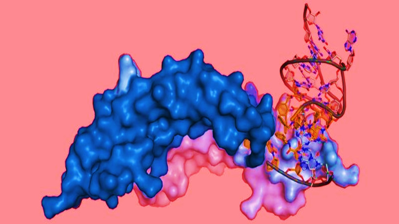
The hydrogel at the heart of this biosensor is constructed from cross-linked acrylamide polymers functionalized with DNA strands. These strands provide both structural integrity and selective recognition capacity, since their complementary sequences interact with target miRNAs. When no target is present, the hydrogel maintains a three-dimensional porous matrix that physically traps HRP molecules inside. The confinement prevents enzyme leakage, maintaining assay stability and minimizing background noise in measurements. Scanning electron microscopy reveals this architecture as a mesh-like network, while spectroscopic analysis confirms HRP encapsulation. Thus, the hydrogel serves a dual role: structural reservoir and responsive actuator.
Upon introduction of a target miRNA, hybridization with the linker DNA sequence destabilizes the hydrogel. This collapse event is not merely structural but functional, as it results in the liberation of HRP into the surrounding medium. Once released, the enzyme encounters its substrate, 3,3′,5,5′-tetramethylbenzidine (TMB), in the presence of hydrogen peroxide. HRP catalyzes the one-electron oxidation of TMB, generating a blue-colored oxidized product with distinct optical absorbance. This oxidation product is photothermally active, absorbing NIR light and converting it into localized heat. Consequently, a molecular recognition event is amplified into a macroscopic physical effect.
The biophysical properties of the hydrogel are fine-tuned to optimize sensitivity and specificity. Factors such as cross-linking density, enzyme loading concentration, and hybridization temperature all determine system performance. For example, overly dense cross-linking hinders enzymatic release, while insufficient cross-linking risks premature leakage. Optimal loading of HRP balances sensitivity with reproducibility, ensuring that temperature readouts reflect genuine target interactions. Moreover, pH conditions are calibrated to maintain HRP activity while avoiding nonspecific oxidation events. These engineering considerations exemplify the delicate interplay between polymer chemistry, molecular recognition, and enzymatic kinetics.
Critically, hydrogels are inherently biocompatible and adaptable to various modifications. By altering the DNA linker sequences, one can design hydrogels responsive to different miRNAs or even broader classes of nucleic acids. This modularity suggests the platform can evolve into a versatile diagnostic toolkit rather than a single-use assay. The hydrogel’s responsiveness also allows integration with multiple readout modalities, expanding its utility beyond thermal detection. These features situate the hydrogel system within the larger movement toward customizable biosensors tailored to specific biomedical contexts. The architecture thus becomes not just a medium for one biomarker but a generalizable framework.
Photothermal Signal Conversion
The innovation of this system lies in translating biochemical recognition into thermal signatures. Oxidized TMB exhibits strong absorption in the near-infrared region, enabling efficient conversion of light into heat. When irradiated with an 808 nm laser, localized heating occurs within seconds, creating measurable temperature elevations. This effect provides a direct, quantitative correlate to the amount of HRP released, which in turn reflects miRNA abundance. Importantly, the heating process is reversible and non-destructive, allowing repeated measurements without compromising sample integrity. Such stability makes photothermal conversion especially appealing for clinical contexts requiring consistency.
The photothermal principle resolves longstanding limitations in optical biosensing. Traditional absorbance-based systems depend on spectrophotometers, which are costly and immobile. In contrast, temperature measurement can be accomplished with widely available digital thermometers, which are inexpensive and portable. This democratizes access to advanced molecular diagnostics, particularly in low-resource settings. Additionally, thermal readouts are robust against environmental fluctuations that often confound optical or electrochemical signals. The system therefore enhances reliability while lowering operational barriers.
Photothermal amplification is inherently linear across a wide concentration range, which aligns well with diagnostic needs. Incremental increases in miRNA concentration lead to proportional rises in temperature under controlled irradiation conditions. This linearity simplifies calibration, avoiding the nonlinear saturation effects often observed in fluorescence-based systems. Moreover, the low detection threshold demonstrates that the thermal method retains sensitivity comparable to PCR-based assays. Such equivalence underscores the biosensor’s potential as a point-of-care surrogate for gold-standard molecular diagnostics. Thermal conversion thus becomes not only a readout mechanism but a validation of molecular recognition fidelity.
Beyond diagnostic use, photothermal detection has implications for therapeutic monitoring. Since miRNAs are implicated in drug resistance and tumor microenvironment modulation, real-time quantification could inform treatment strategies. A thermometer-based approach could be deployed during chemotherapy to track circulating miRNA levels as a proxy for tumor response. Furthermore, the underlying principle could extend to monitoring other nucleic acids or proteins that generate photothermal signatures upon enzymatic conversion. This adaptability situates photothermal biosensing within broader personalized medicine initiatives. Hence, temperature emerges as both a literal and metaphorical indicator of biological activity.
Optimization and Performance Evaluation
Systematic optimization was required to ensure robust biosensor performance. Researchers evaluated incubation times to establish equilibrium between miRNA binding and hydrogel collapse, identifying two hours as sufficient for complete reaction. Enzyme concentration was titrated to prevent leakage while preserving catalytic efficiency, with four micrograms per milliliter determined optimal. Hydrogen peroxide levels were carefully balanced, since excess reagent inhibited HRP activity by promoting oxidative stress. Temperature and pH ranges were also refined, with 30°C and pH 4.0 yielding maximal catalytic turnover. These parameters collectively define the operational envelope of the biosensor.
Performance testing confirmed sensitivity, selectivity, and reproducibility across multiple assays. Increasing concentrations of miR-21 from HeLa cells produced consistent shifts from colorless to blue, easily perceptible to the naked eye. Corresponding temperature rises under NIR illumination exhibited strong linear correlation with miRNA levels, reinforcing the assay’s quantitative capacity. Repeat experiments demonstrated stability over several weeks, indicating that both hydrogel integrity and enzymatic activity were preserved. Selectivity tests using unrelated miRNAs confirmed that the hydrogel linker sequences imparted specificity through complementary hybridization. These outcomes collectively highlight the system’s clinical readiness.
The biosensor’s reproducibility is particularly notable, as enzymatic assays often suffer from variability. By embedding HRP within the hydrogel rather than in free solution, catalytic consistency was maintained across trials. This encapsulation strategy reduces enzyme denaturation and shields against environmental fluctuations. Furthermore, the three-dimensional hydrogel network regulates diffusion kinetics, ensuring uniform exposure of HRP to substrates upon release. Such control enhances both signal fidelity and assay longevity. These structural advantages distinguish the hydrogel-based system from conventional liquid-phase assays.
Transitioning from bench to bedside requires validation with real biological samples. The biosensor was tested with miRNA extracted directly from HeLa cell lysates and compared against qRT-PCR, the current gold standard. Results demonstrated near-identical detection profiles, confirming the biosensor’s analytical accuracy. This equivalence provides confidence that portable, thermometer-based detection can rival sophisticated laboratory methods. In practical terms, the assay can be deployed in contexts where PCR infrastructure is inaccessible. Such validation positions the hydrogel biosensor as a credible alternative for clinical application.
Toward Portable Molecular Diagnostics
The broader implication of this work is the decentralization of molecular diagnostics. By eliminating dependence on specialized laboratory equipment, the hydrogel biosensor democratizes access to advanced testing. Portable thermometer readouts transform what is typically a resource-intensive process into a universally approachable tool. This accessibility aligns with global health priorities that emphasize equitable distribution of diagnostic capability. The biosensor’s simplicity also reduces operator error, further enhancing reliability in diverse contexts. In this way, engineering choices intersect directly with public health imperatives.
The modularity of DNA hydrogel systems expands the diagnostic landscape. Linker sequences can be tailored to recognize distinct miRNAs associated with various cancers or infectious diseases. This adaptability means a single platform could address multiple biomarkers simply by altering crosslinking DNA. Furthermore, the photothermal principle could integrate with smartphone-based temperature sensors, linking results directly to digital health infrastructures. Such convergence of biotechnology and mobile technology heralds a new era of distributed diagnostics. The biosensor thus serves as both a scientific innovation and a technological bridge.
Integration into clinical workflows requires attention to scalability and manufacturability. Hydrogels must be produced consistently and stored without losing functional responsiveness. Enzyme encapsulation techniques will need industrial refinement to support commercial deployment. Packaging assays into standardized, single-use kits could accelerate adoption in hospitals and clinics. Regulatory validation will also be necessary to establish equivalence with established PCR methodologies. These steps mark the path from experimental validation to widespread adoption.
Ultimately, the HRP-encapsulated DNA hydrogel biosensor represents a paradigm shift in biomolecular detection. It transforms the abstract information of miRNA sequences into tangible, measurable heat. The portability of a thermometer as a detection tool lowers barriers to entry without compromising scientific precision. As healthcare systems continue to embrace personalized medicine, such innovations will become indispensable in early detection and monitoring. This biosensor is therefore not merely a diagnostic curiosity but a glimpse into the future of molecular medicine.
Study DOI: https://doi.org/10.3389/fbioe.2021.799370
Engr. Dex Marco Tiu Guibelondo, B.Sc. Pharm, R.Ph., B.Sc. CpE
Editor-in-Chief, PharmaFEATURES

Subscribe
to get our
LATEST NEWS
Related Posts
Molecular Biology & Biotechnology
Aptamer Targeting Nanomedicine: Programmable Nanocarriers for Precise, Multi-Organ Tumor Therapy
Programmable aptamer–nanocarrier systems turn the tumor microenvironment into an addressable code, enabling precise, multi-modal therapy with real-time control.

Molecular Biology & Biotechnology
Intracellular Arsenal: Cell Permeating Nanomaterials Eliminating Bacteria via Targeted Delivery and Intrinsic Bioactivity
Engineered nanomaterials transform intracellular infection therapy by synchronizing targeted delivery with built-in antimicrobial bioactivity inside the very compartments bacteria use to hide.
Molecular Biology & Biotechnology
Microarchitects of Fertility: How Sertoli Cells Script Germline Renewal
Sertoli cells engineer male fertility by integrating paracrine signals, juxtacrine contact, and epigenetic regulation within the spermatogonial stem cell niche.

Molecular Biology & Biotechnology
Cord Hemoglobin Redux: Ascorbate-Stabilized Polymerized Fetal Hemoglobin Tames Renal Oxidative Injury During Oxygen-Bridge Transfusion
Pairing polymerized fetal hemoglobin with ascorbate converts a fragile acellular oxygen carrier into a clinically plausible, kidney-aware oxygen-bridging therapy.
Read More Articles


Medicinal Chemistry & Pharmacology
April 14, 2026
Igor Nasonkin and Phythera Therapeutics: Moving Oncology Beyond Single Targets into Engineered Polypharmacologic Systems
Igor Nasonkin’s systems-driven approach at Phythera Therapeutics reframes oncology drug development from single-target inhibition to AI-enabled polypharmacologic network modulation using nature-derived molecular architectures.


Artificial Intelligence and Data Analytics
April 10, 2026
Inside Johnson & Johnson’s External Innovation Engine: Devin Swanson on Translating Integrated Discovery into Strategic Value
Devin Swanson’s leadership at Johnson & Johnson Innovative Medicines redefines external innovation as a tightly governed, AI-enabled translational system integrating multi-modal drug discovery, biomarker strategy, and capital-efficient execution.

Immunology & Oncology
April 9, 2026
From DMPK to Distributed Execution: Mehran F. Moghaddam’s Systems Strategy at OROX BioSciences, Inc.
A systems-level examination of how Mehran F. Moghaddam operationalizes DMPK, externalized R&D, and lipid-mediated therapeutics into a predictive, high-velocity biotech development architecture.