Chitosan-Salvianolic Acid B Complex on Nickel-Titanium Alloys as a Blueprint for Endothelialization and Anti-Restenosis Strategies
Rethinking the Biology of Stent Surfaces
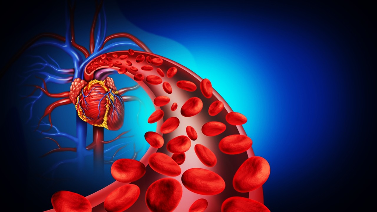
The clinical deployment of intracranial stents has transformed how cerebrovascular disease is managed, providing structural scaffolds to reopen narrowed vessels. Yet, the longevity of these implants has consistently been hindered by two opposing biological imperatives: suppressing the unchecked proliferation of vascular smooth muscle cells while simultaneously encouraging the re-establishment of an intact endothelial layer. Drug-eluting stents were once hailed as the definitive solution, but their legacy has been clouded by complications associated with delayed re-endothelialization, chronic inflammation, and long-term thrombosis.
At the molecular level, the vascular wall functions as a delicate equilibrium between repair and remodeling. Smooth muscle cells, which proliferate aggressively after vessel injury, form the basis of restenosis. Endothelial cells, in contrast, regulate hemostasis, vascular tone, and immune interactions, and their absence transforms the stented lumen into a thrombogenic surface. This duality has long challenged material scientists, pharmacologists, and clinicians: how can a single surface chemistry direct two distinct cellular fates?
Nickel-titanium alloys, celebrated for their shape memory and mechanical resilience, have served as an ideal scaffold for intracranial and peripheral stents. However, the alloy itself is biologically inert and prone to protein adsorption, coagulation activation, and cellular misrecognition. Hence, modifying its surface with bioactive coatings has become the frontier of vascular implant science. The introduction of biofunctional interfaces that integrate drug delivery and biomimetic cues now represents a significant evolution beyond polymer-only stents.
The current line of inquiry lies in engineering multifunctional coatings that can release therapeutic molecules with temporal precision while simultaneously altering surface hydrophilicity, electrostatic interactions, and adhesion kinetics. Among the promising strategies is the immobilization of salvianolic acid B—an antioxidant phenolic compound derived from Salvia miltiorrhiza—within a chitosan polymeric matrix applied onto nickel-titanium alloys. The rationale is strikingly simple yet profoundly effective: pair a vascular-protective phytochemical with a biodegradable delivery scaffold to redefine how stent coatings interact with blood and vascular cells.
Salvianolic Acid B: A Molecular Bridge Between Repair and Restraint
Salvianolic acid B (SALB) is not a synthetic pharmacological invention but rather a naturally occurring small molecule that has co-evolved within traditional medicinal practices. Its structure, a caffeic acid trimer, underpins strong antioxidant potential while also modulating signaling cascades relevant to vascular pathology. In preclinical contexts, SALB has shown the ability to suppress reactive oxygen species, inhibit inflammatory mediators, and modulate apoptotic pathways. These attributes extend beyond general cytoprotection; they directly intersect with the molecular mechanisms that govern endothelial cell survival and smooth muscle proliferation.
When endothelial cells encounter SALB, there is a documented upregulation of pro-angiogenic markers, including vascular endothelial growth factor receptors and phosphorylated STAT3. This upregulation translates into improved endothelial migration and adhesion, critical events in re-establishing the vascular lining post-stenting. Simultaneously, SALB has demonstrated the capacity to interfere with smooth muscle cell cycle regulators and signaling nodes such as CXCR4, effectively dampening their proliferative drive. The duality of this molecular profile positions SALB as uniquely capable of resolving the paradox of stent biology: fostering re-endothelialization while preventing neointimal hyperplasia.
Yet, SALB is not without limitations. Its pharmacokinetic profile reveals a relatively short half-life and rapid clearance, characteristics that undermine its sustained action in vivo. For a stent coating, this pharmacological transience could reduce its therapeutic window and limit its clinical utility. Thus, material scientists have turned to polymeric carriers such as chitosan to encapsulate SALB and extend its release over clinically relevant timescales.
This marriage of bioactive compound and biopolymer provides more than pharmacokinetic modulation. Chitosan, a linear polysaccharide derived from chitin, is inherently biocompatible, biodegradable, and structurally versatile. Its positively charged amine groups interact electrostatically with biomolecules and cellular membranes, making it an effective mediator of cell-material interactions. By entrapping SALB in chitosan microspheres, researchers create a reservoir system where drug release occurs in two phases: an early burst and a prolonged slow-release phase governed by polymer degradation.
Thus, the introduction of SALB into vascular stent coatings exemplifies a convergence of natural product pharmacology and polymer engineering. It is not merely about attaching a molecule to a surface but about orchestrating a complex interplay between cellular responses, surface chemistry, and sustained bioactivity.
Chitosan as a Vehicle for Controlled Release and Biocompatibility
Chitosan’s value in biomedical engineering transcends its role as a passive carrier. As a polysaccharide composed primarily of glucosamine units, its structural features—degree of deacetylation, molecular weight, and chain length—dictate solubility, degradation kinetics, and biological reactivity. These variables can be tuned to optimize drug encapsulation, loading efficiency, and release behavior. In the context of SALB delivery, chitosan nanoparticles are prepared through ionic crosslinking with sodium tripolyphosphate, which introduces electrostatic stability without resorting to harsh chemical crosslinkers.
The resultant microspheres are capable of entrapping SALB while maintaining its structural integrity. Once immobilized onto a polydopamine-coated nickel-titanium surface, the chitosan-SALB matrix adheres through both physical adsorption and covalent interactions, minimizing the risk of detachment under hemodynamic shear. Moreover, chitosan modifies the hydrophilicity of the metallic surface, enhancing wettability and thereby influencing protein adsorption kinetics. Surfaces with intermediate hydrophilicity, as observed in chitosan-modified alloys, have been shown to support more physiologic endothelial adhesion patterns.
In vitro drug release studies confirm that the chitosan-SALB complex avoids the pitfall of rapid drug depletion. Instead, release occurs steadily over a period measured in weeks, a timeframe aligned with the critical window of endothelial recovery following stent implantation. The controlled degradation of chitosan ensures a continuous supply of SALB molecules to the peri-stent tissue, maintaining biological pressure against smooth muscle proliferation while promoting endothelial restitution.
Beyond release dynamics, chitosan introduces intrinsic biological activities to the coating. It possesses mild antimicrobial effects, limiting bacterial adhesion in the early post-implantation period. Its degradation products are non-toxic and metabolizable, reducing long-term inflammatory sequelae. Moreover, chitosan’s amine-rich backbone interacts favorably with plasma proteins, reducing platelet adhesion and lowering the thrombogenic profile of the stent. These converging benefits transform chitosan from a mere vehicle to a co-therapeutic component of the stent coating.
The utilization of chitosan in this context is emblematic of the broader shift in biomaterials research: toward multifunctional scaffolds that combine mechanical stability with biological intelligence. By linking drug delivery with structural modification of the implant surface, chitosan enhances not only pharmacological efficacy but also cellular compatibility.
Biological Outcomes: Endothelialization and Suppression of Restenosis
The dual performance of the chitosan-SALB coating has been validated in vitro through carefully designed cellular assays. Human umbilical vein endothelial cells cultured on coated surfaces exhibit accelerated adhesion, migration, and proliferation compared to bare alloy controls. This suggests that the surface chemistry of the composite coating creates a microenvironment that mimics the extracellular matrix cues necessary for endothelial recovery. Fluorescence imaging further reveals well-spread endothelial morphologies, polygonal in shape, indicating cytoskeletal organization conducive to barrier function.
Smooth muscle cells respond in a markedly different manner. Their adhesion to chitosan-SALB surfaces is reduced, and their morphology becomes spindle-like and narrowed, reflecting a cytoskeletal adaptation associated with reduced proliferation. Migration assays confirm that the coating impedes smooth muscle invasiveness, effectively limiting the cellular cascade that culminates in restenosis. This selective cellular modulation underscores the precision of the dual-function design.
Blood compatibility, another cornerstone of implant success, is equally improved. Protein adsorption assays show reduced fibrinogen deposition, a critical factor in platelet activation and thrombus initiation. Coagulation assays further reveal extended clotting times, suggesting that the modified surfaces attenuate pro-coagulant triggers. Collectively, these outcomes present a surface that is not only structurally supportive but also dynamically engaged with blood elements in a way that mitigates thrombosis.
From a translational perspective, these cellular and biochemical findings address the two most persistent clinical challenges of intracranial stenting: neointimal hyperplasia and thrombosis. By directing endothelial recovery while restraining smooth muscle pathology, the chitosan-SALB coating redefines what can be expected from drug-eluting stent technology. The implication is not incremental improvement but rather a paradigm shift in how implantable devices interact with living tissues.
Toward Clinical Translation and Future Directions
The promise of chitosan-SALB coatings lies not only in their mechanistic plausibility but also in their compatibility with scalable manufacturing processes. Polydopamine-assisted immobilization of polymer-drug microspheres can be applied to commercial stents using existing surface modification infrastructure. This facilitates a realistic pathway from laboratory alloy plates to medical-grade devices without fundamental redesign of stent platforms.
Nevertheless, challenges remain before clinical deployment. In vivo testing across animal models is essential to assess long-term biocompatibility under hemodynamic conditions. Endothelial responses observed in vitro must be validated in the complex milieu of vascular injury, inflammation, and systemic circulation. Furthermore, questions regarding dose optimization, degradation kinetics, and patient variability in drug metabolism will require comprehensive pharmacological studies.
Another avenue of inquiry involves dissecting the precise molecular signaling networks modulated by SALB. While current evidence points to VEGF and STAT3 pathways in endothelial cells and cell cycle regulators in smooth muscle cells, the broader genomic and proteomic landscapes remain to be mapped. Understanding these interactions could enable further refinement of the coating, such as combining SALB with additional bioactive agents for synergistic effects.
Beyond cerebrovascular interventions, the implications of this coating extend to peripheral and coronary stents, vascular grafts, and even non-vascular implants where dual control of cellular adhesion is required. The chitosan-SALB complex represents a modular approach that can be tailored to different clinical contexts by adjusting drug loading, microsphere size, or coating density.
The future of stent technology lies in precisely engineered biointerfaces that are as biologically intelligent as they are mechanically robust. The chitosan-SALB coating exemplifies this vision, offering a surface that not only passively resides in the vasculature but actively participates in the orchestration of healing. As research progresses, this dual-action design may become the blueprint for next-generation vascular implants.
Study DOI: https://doi.org/10.3389/fbioe.2023.1300336
Engr. Dex Marco Tiu Guibelondo, B.Sc. Pharm, R.Ph., B.Sc. CpE
Editor-in-Chief, PharmaFEATURES

Subscribe
to get our
LATEST NEWS
Related Posts
Regenerative Medicine & Biomaterials
Programmable Nitric Grafts: Steady Nitric Oxide Orchestrates Small-Vessel Healing
Embedding nitrate-driven nitric-oxide release within PCL grafts programmatically guides vascular regeneration across the lumen, media, and progenitor niche.

Regenerative Medicine & Biomaterials
Bioactive Networks: Photocrosslinkable Gelatin Hydrogels and Cytokine Modulation in Human Immunity
GelMA hydrogels demonstrate that the architecture of matter itself can modulate the architecture of inflammation.

Read More Articles


Medicinal Chemistry & Pharmacology
April 14, 2026
Igor Nasonkin and Phythera Therapeutics: Moving Oncology Beyond Single Targets into Engineered Polypharmacologic Systems
Igor Nasonkin’s systems-driven approach at Phythera Therapeutics reframes oncology drug development from single-target inhibition to AI-enabled polypharmacologic network modulation using nature-derived molecular architectures.


Artificial Intelligence and Data Analytics
April 10, 2026
Inside Johnson & Johnson’s External Innovation Engine: Devin Swanson on Translating Integrated Discovery into Strategic Value
Devin Swanson’s leadership at Johnson & Johnson Innovative Medicines redefines external innovation as a tightly governed, AI-enabled translational system integrating multi-modal drug discovery, biomarker strategy, and capital-efficient execution.

Immunology & Oncology
April 9, 2026
From DMPK to Distributed Execution: Mehran F. Moghaddam’s Systems Strategy at OROX BioSciences, Inc.
A systems-level examination of how Mehran F. Moghaddam operationalizes DMPK, externalized R&D, and lipid-mediated therapeutics into a predictive, high-velocity biotech development architecture.