From Intertidal Adhesion to Microbial Targeting: The Polydopamine Interface
Mussel foot proteins solved a hard materials problem first: how to adhere wet, charged, and rough surfaces without primers, and that chemical grammar seeded an entire surface chemistry. Polydopamine, the synthetic echo of that grammar, forms in mildly alkaline buffers and blankets disparate substrates with a thin, catechol- and amine-rich film. Those catechols and amines participate in hydrogen bonding, π–π stacking, Schiff base formation, and Michael addition, giving one coating many orthogonal handles for bioconjugation. In bioengineering, that universality has been repeatedly exploited to immobilize peptides, polysaccharides, and small molecules under gentle conditions compatible with live cells. A mussel-mimetic shell therefore becomes a logical chassis for nanoprobes that must both survive complex media and present selective ligands. That is the conceptual leap behind polydopamine-coated silicon quantum dots for Gram-aware labeling of bacteria and their biofilms.
When polydopamine forms around a colloidal core, it functions as more than glue; it is a chemical switchboard. The coating throttles non-specific interactions through tunable charge and hydrophilicity while simultaneously exposing motifs that can be clicked, stacked, or ion-paired. In the context of microbial imaging, this duality is critical because cell envelopes differ in peptidoglycan organization, lipopolysaccharide content, and surface charge distribution. One needs a scaffold that neither collapses in salt nor fouls in proteinaceous media yet can bias interactions toward a chosen envelope target. Polydopamine’s supramolecular promiscuity is not a bug but a design feature, allowing the same nanoprobe to be reprogrammed by swapping a small molecule at the interface. That versatility underwrites the selective probes described here.
Selectivity at the nanoscale is a subtle balance between affinity and access through the envelope’s barriers. Gram-positive species present thick, porous peptidoglycan with teichoic acids, while Gram-negative species present an asymmetric outer membrane with lipopolysaccharide acting as a kinetic and electrostatic gate. A single nanostructure cannot simply be labeled “universal” unless its interface can be retuned to those distinct physicochemical landscapes. Polydopamine allows that retuning by supporting ligand exchange and multivalent display without compromising colloidal stability. The result is a platform where chemistry, not the core, dictates targeting logic. In practice, that means the same quantum dot can be made Gram-positive-seeking one day and Gram-negative-seeking the next.
These design aims matter because traditional labeling workflows carry practical burdens. Gram staining is discontinuous, destructive, and population-averaging, while nucleic-acid-centric assays demand sample prep and equipment that are not always fieldable. Fluorogenic organic dyes bring speed but often photobleach under illumination regimes needed for time-lapse or thick-sample imaging. Semiconductor nanocrystals can resist that bleaching but raise concerns about heavy-metal content and cytotoxicity. Silicon quantum dots, by contrast, offer bright, narrow emission and benign composition, positioning them as photophysically robust yet biologically palatable cores. Marrying them to a mussel-inspired shell closes the loop between materials resilience and biochemical addressability.
Engineering a Core–Shell Nanoprobe: Green-Emissive SiQDs Under an Imitation-Mussel Shell
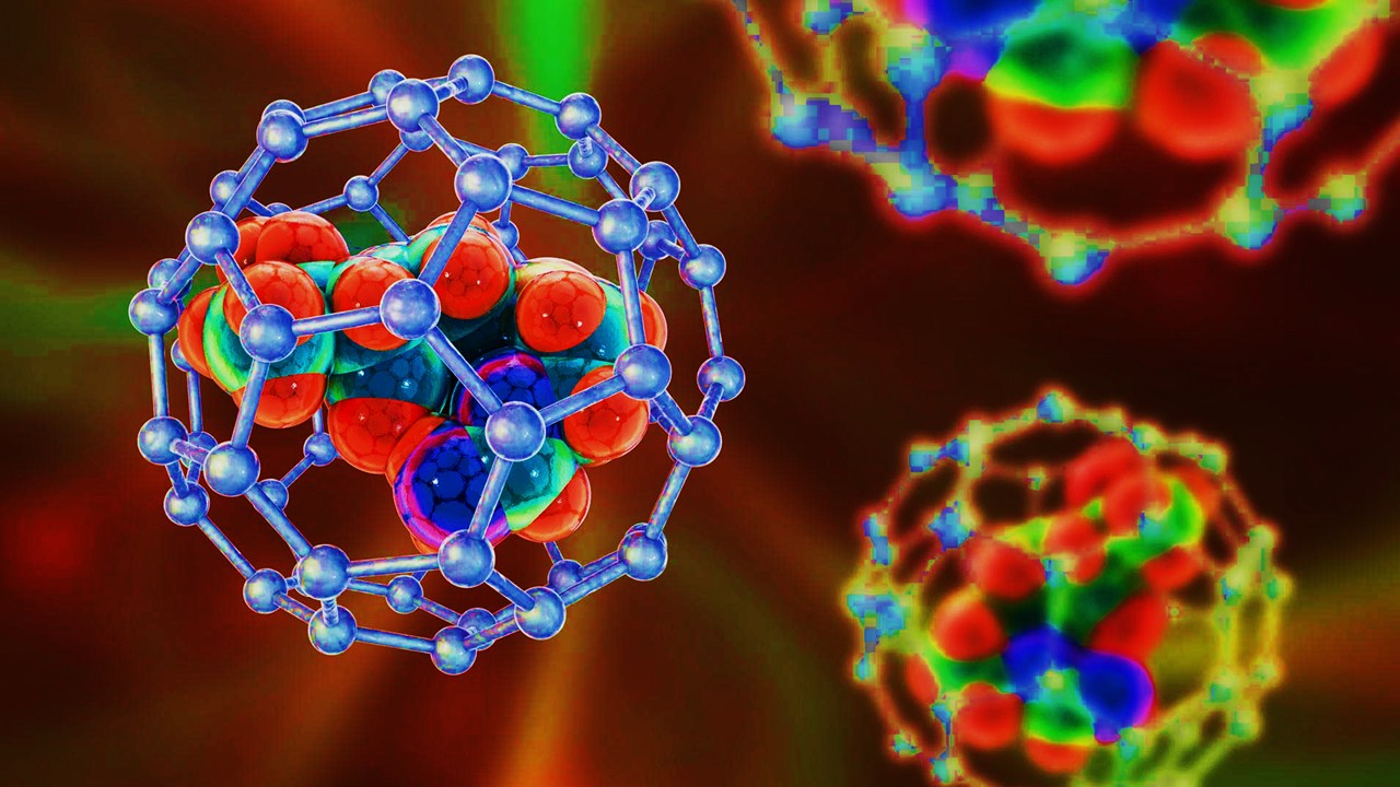
The silicon cores are prepared from aminosilanes in water under a dye-sensitized hydrothermal route, yielding ultrasmall, green-emissive quantum dots that disperse cleanly. Transmission electron microscopy shows isolated nanocrystals with diameters in the single-nanometer regime, consistent with strong quantum confinement. Spectroscopy places the excitation in the blue-green and emission in the green, aligning with standard FITC channels for straightforward microscopy integration. X-ray diffraction indicates an amorphous or poorly crystalline silicon framework, a common feature in solution-grown SiQDs that nonetheless supports bright photoluminescence. The critical observation is that the optical band structure survives shelling, preserving usable brightness for live imaging. This is the core photophysical footing on which the biological story stands.
Polydopamine is then deposited in Tris buffer at mild pH, where dopamine oxidizes and polymerizes to form a conformal shell around the silicon core. Electron micrographs reveal a growth in hydrodynamic size consistent with a thin organic layer, and Fourier-transform infrared spectra show the emergence of aromatic and amine signatures attributable to the polydopamine network. Ultraviolet–visible measurements show that the shell overlays the core’s absorption features without abolishing the emissive transition. The small drop in observed intensity is offset by gains in chemical robustness and functionalization capacity afforded by the coating. Colloidal stability remains intact, with no aggregation observed under the imaging conditions reported. The shell, in effect, trades a fractional quantum yield penalty for a significant leap in biochemical programmability.
Functionality at the shell is validated by three orthogonal conjugation modes that map to polydopamine’s chemistry. Aromatic antibiotics couple via π–π interactions to the catechol/indole-like domains, cationic amino acids adhere via ion–ion contacts to the negatively charged surface, and thiol-bearing ligands add across quinone-like vinyls in a Michael addition. Infrared spectroscopy tracks characteristic carbonyl and hydroxyl bands from each ligand class after conjugation, while X-ray photoelectron spectroscopy reports shifts in elemental ratios consistent with new organic content. Zeta potential shifts corroborate successful surface exchanges when the ligand introduces strong charge asymmetry. The upshot is universal functionalization under one mild recipe rather than bespoke silanization for every ligand. That universality is precisely what a practical microbial probe requires.
Photophysical durability is a differentiator once the probe meets the microscope. Under continuous white-light illumination, green organic nucleic-acid dyes typically exhibit rapid decay in emission, limiting time-resolved imaging of slow-forming biofilms. The silicon-core, polydopamine-shelled probes sustain their signal substantially longer under the same conditions, enabling side-by-side comparisons without frequent replenishment or exposure minimization tricks. This resilience arises from the inorganic core’s resistance to photochemical degradation and the shell’s role in quenching surface traps. Practically, that means less signal drift across a z-stack and more reliable quantitation across replicates. It also means fewer phototoxicity confounders from high excitation powers used to compensate for fading dyes. The material choice thus directly maps to data quality in living samples.
Wiring Molecular Recognition: D-alanine for Peptidoglycan, Gluconamide for Lipopolysaccharide
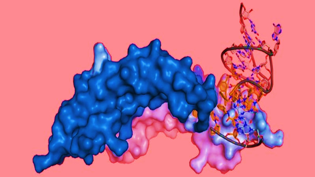
The selectivity mechanism is built from bacterial biochemistry rather than antibody epitopes. D-alanine is a canonical component of the peptidoglycan stem peptide and cycles through transpeptidases and ligases during cell-wall synthesis, allowing exogenous D-alanine derivatives to report on or ride into the wall. When tethered to the polydopamine shell, D-alanine biases the nanoprobe toward envelopes where peptidoglycan is thick and accessible, as in Gram-positive organisms. This is not simply electrostatics but a metabolism-aware interaction with precursors and enzymes at the wall. Fluorescent D-amino acid labeling has previously visualized growth patterns and envelope remodeling, and the present design leverages that biochemical doorway on a nanoparticle scaffold. The core–shell architecture therefore translates a metabolic handle into an imaging handle.
Gluconamide provides a complementary handle for Gram-negative targeting by engaging lipopolysaccharide at the outer membrane. Molecular simulations and experimental studies have shown that gluconamide groups insert and interact favorably within the lipopolysaccharide region, providing proximity to the outer leaflet without harsh permeabilization. Displayed multivalently on a polydopamine shell, these groups can achieve short-range, carbohydrate-mediated interactions at the membrane surface. Such interactions thread a path through the barrier properties that normally frustrate probe access to Gram-negative cells. Because lipopolysaccharide composition is conserved in its architectural features even as chemotypes vary, the approach generalizes across species. The ligand choice thus encodes the Gram logic directly into the nanoprobe.
Putting the two ligands on identical cores isolates the variable that matters: the interface. Surface characterization confirms successful D-alanine and gluconamide installation via appearance of ligand-specific vibrational bands and shifts in elemental ratios. Control conjugations using small thiols and PEG chains further demonstrate that the Michael addition route is agnostic to molecular size over orders of magnitude. Zeta potential data provide an orthogonal readout that, while moderated by hydroxyl-rich ligands, reflects the new corona chemistry. The point is not to create a heavily charged particle but to present the right motifs to the right biology. In that sense, polydopamine acts as a programmable transducer between inorganic photophysics and microbial surface biochemistry.
This recognition logic suggests further engineering trajectories beyond single-color labels. Ratiometric constructs, where a reference emission remains constant while a ligand-gated channel changes, could deconvolve binding from local concentration. Orthogonal ligand sets could support multiplex Gram and genus-level panels in the same field of view. Swapping the emission into the red or near-infrared would reduce background and improve tissue penetration for ex vivo specimens. Incorporating cleavable linkers sensitive to wall-associated enzymes might convert binding into catalytic signal gain. Each of these variations keeps the polydopamine interface but edits how and what it displays. The scaffold is thus a platform rather than a one-off probe.
Gram-Specific Labeling in Planktonic Cells and 3D Biofilms
With D-alanine on board, the probe lights up Gram-positive cells under standard fluorescence microscopy, producing bright signal in Staphylococcus aureus and Enterococcus faecalis while leaving Gram-negative controls near background. Flow cytometry corroborates the imaging, shifting fluorescence distributions for the targeted species relative to untreated controls. Variability among Gram-positive species appears consistent with known differences in peptidoglycan architecture and accessibility. The important feature is that labeling emerges without permeabilization or wash-intensive workflows that perturb physiology. Conversely, the gluconamide-functionalized probe shows labeling of Escherichia coli and Pseudomonas aeruginosa and minimal signal in Gram-positive comparators. The two sister constructs therefore function as a simple Gram test deployable on a microscope.
Biofilms bring their own physical chemistry, with extracellular polymeric substances modulating diffusion, adsorption, and local pH. In printed three-dimensional biofilms, the D-alanine probe penetrates and illuminates Gram-positive structures, while the gluconamide probe does the same in Gram-negative constructs. Imaging reveals contiguous fluorescent architectures rather than only peripheral staining, indicating access through the hydrogel-like matrix. Because both probes emit in the green, acquisition parameters mirror those used for common live-dead assays, simplifying adoption. The shell’s hydrophilicity and ligand presentation likely mitigate non-specific trapping in the polysaccharide mesh. The outcome is readable contrast inside living, architected consortia rather than only in planktonic cultures.
Under prolonged illumination, the silicon-core probes resist the rapid fading that constrains many organic dyes in biofilm imaging. That stability maintains signal over the time windows needed to follow attachment, microcolony maturation, and matrix deposition. In comparative measurements, the organic green nuclear dye drops steeply, while the quantum-dot signal declines far more gently under identical lighting. For practitioners, that translates into fewer exposure adjustments, reduced normalization burdens, and better comparability across timepoints. It also enables extended depth scans in thick samples without losing the channel mid-stack. Photophysical robustness thus becomes a biological advantage in long-form imaging.
Cytocompatibility complements photostability because labeling should not alter growth dynamics in the observation window. Assays on mammalian cell lines indicate minimal acute toxicity at working concentrations for the core, the shelled dot, and both ligand-functionalized variants. That profile aligns with the literature view of silicon quantum dots as a safer alternative to heavy-metal nanocrystals when intended for biological contact. It further benefits from polydopamine’s wide biomedical use, where coatings have been shown to be compatible with diverse cell types. While comprehensive toxicology in complex in vivo settings remains future work, the current data justify use in in vitro microbiology and ex vivo imaging. The materials decisions are thus compatible with the experimental ethics of live-cell microscopy.
Translating Photophysics Into Practice: Assay Design, Multiplexing, and Device Integration
For laboratories, the immediate utility is a fast, wash-light, Gram-aware label that slots into existing fluorescence microscopes and flow cytometers. Sample prep consists of co-incubation and gentle rinses, and analysis can be scripted using standard FITC-channel workflows. Because the interface chemistry is modular, one can imagine kit-style vials where the same stock SiQDs@PDA is derivatized on demand with a chosen ligand. That reduces inventory complexity and supports side-by-side comparisons without batch-to-batch spectral drift. The platform also invites integration with microfluidic bacterial culture devices, where laminar co-flows can deliver distinct probes for rapid phenotyping. In microbial ecology and clinical triage alike, that simplicity can accelerate actionable readouts.
Looking forward, the Gram logic demonstrated here is a template rather than a terminus. Ligands that read teichoic-acid patterns, porin repertoires, or membrane potential could be grafted to build higher-resolution phenotypic panels. Enzyme-responsive linkers could report on β-lactamase activity or periplasmic redox state, coupling labeling to function. Shifting emission into the red or near-infrared would improve penetration and reduce autofluorescence in tissue sections or thick biofilms. Multiplexing cores with distinct emissions on the same shell could enable barcoded probes for community-level mapping. Each variant would still rely on the polydopamine switchboard to wire recognition chemistry to a bright, stable core. The strategy scales because the interface scales.
Clinical and industrial microbiology pose additional constraints that the platform is well positioned to meet. Stability in transport media, compatibility with automated staining stations, and resilience under varied illumination conditions are non-negotiables outside research settings. Silicon cores and polydopamine shells are inherently suited to those demands, and ligand choice can be tuned to target contaminants or pathogens relevant to the process line. In environmental monitoring, for example, membrane-targeting ligands could flag Gram-negative contamination in water or bioprocess streams with minimal sample preparation. In infectious disease workflows, Gram-aware labeling upstream of culture could shorten decision cycles for empirical therapy. The business case thus rides on the same material choices that drove the science.
No platform is complete without acknowledging failure modes and edge cases. Thick capsules and surface polysaccharide diversity in some species may reduce access for both probes, motivating accessory permeabilization-free strategies. Biofilm heterogeneity can generate locally acidic or oxidizing microenvironments that perturb polydopamine chemistry over long incubations. Competitive binding from serum proteins or extracellular DNA may dampen signal in complex matrices, suggesting the value of antifouling co-polymers alongside the active ligand. Photophysics can also shift subtly with shell thickness, calling for batch-level calibration curves when quantitative intensity is required. These are solvable engineering problems rather than fundamental incompatibilities. Addressing them will broaden the domain where mussel-mimetic SiQDs deliver reliable microbial maps.
Study DOI: https://doi.org/10.3389/fbioe.2022.971682
Engr. Dex Marco Tiu Guibelondo, B.Sc. Pharm, R.Ph., B.Sc. CpE
Editor-in-Chief, PharmaFEATURES

Subscribe
to get our
LATEST NEWS
Related Posts
Molecular Biology & Biotechnology
Aptamer Targeting Nanomedicine: Programmable Nanocarriers for Precise, Multi-Organ Tumor Therapy
Programmable aptamer–nanocarrier systems turn the tumor microenvironment into an addressable code, enabling precise, multi-modal therapy with real-time control.
Molecular Biology & Biotechnology
Intracellular Arsenal: Cell Permeating Nanomaterials Eliminating Bacteria via Targeted Delivery and Intrinsic Bioactivity
Engineered nanomaterials transform intracellular infection therapy by synchronizing targeted delivery with built-in antimicrobial bioactivity inside the very compartments bacteria use to hide.
Molecular Biology & Biotechnology
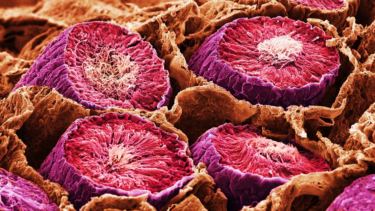
Microarchitects of Fertility: How Sertoli Cells Script Germline Renewal
Sertoli cells engineer male fertility by integrating paracrine signals, juxtacrine contact, and epigenetic regulation within the spermatogonial stem cell niche.
Molecular Biology & Biotechnology
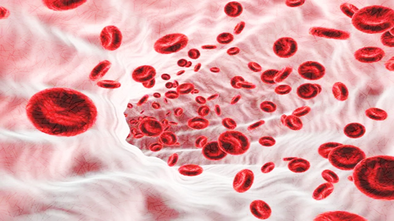
Cord Hemoglobin Redux: Ascorbate-Stabilized Polymerized Fetal Hemoglobin Tames Renal Oxidative Injury During Oxygen-Bridge Transfusion
Pairing polymerized fetal hemoglobin with ascorbate converts a fragile acellular oxygen carrier into a clinically plausible, kidney-aware oxygen-bridging therapy.
Read More Articles


Medicinal Chemistry & Pharmacology
April 14, 2026
Igor Nasonkin and Phythera Therapeutics: Moving Oncology Beyond Single Targets into Engineered Polypharmacologic Systems
Igor Nasonkin’s systems-driven approach at Phythera Therapeutics reframes oncology drug development from single-target inhibition to AI-enabled polypharmacologic network modulation using nature-derived molecular architectures.


Artificial Intelligence and Data Analytics
April 10, 2026
Inside Johnson & Johnson’s External Innovation Engine: Devin Swanson on Translating Integrated Discovery into Strategic Value
Devin Swanson’s leadership at Johnson & Johnson Innovative Medicines redefines external innovation as a tightly governed, AI-enabled translational system integrating multi-modal drug discovery, biomarker strategy, and capital-efficient execution.

Immunology & Oncology
April 9, 2026
From DMPK to Distributed Execution: Mehran F. Moghaddam’s Systems Strategy at OROX BioSciences, Inc.
A systems-level examination of how Mehran F. Moghaddam operationalizes DMPK, externalized R&D, and lipid-mediated therapeutics into a predictive, high-velocity biotech development architecture.