Neurovascular Crosstalk as the Hidden Architecture of Bone Repair
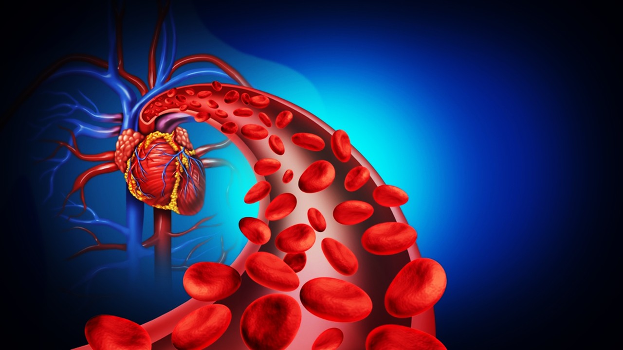
Skeletal regeneration relies not merely on the deposition of minerals but on the synchronized choreography of nerves and vessels within injured tissue. Early frameworks of bone biology emphasized mechanical stress and osteoblast activity, but mounting evidence places neural input and vascular integration at the core of regeneration. In this context, sensory nerves are no longer ancillary messengers of pain but bioactive conduits releasing neuropeptides that reshape osteogenic landscapes. Their interplay with blood vessels defines the tempo of mineralization and ultimately the structural fidelity of new bone. The study of in situ osteogenesis following Achilles tenotomy in rats provides an experimental vantage to dissect this coordinated sequence.
Within this model, sensory nerve fibers first colonize the trauma site, detectable well before calcification thresholds are reached on imaging. These nerves carry calcitonin gene-related peptide and substance P, neuromodulators whose release in bone microenvironments drives local changes in progenitor cell activity. Blood vessels trail behind, their endothelial sprouting often following the contours laid down by early neural infiltration. This spatiotemporal precedence underscores the concept that nerves do not merely accompany healing—they are architects guiding subsequent vascularization. Such precedence raises a fundamental question: which molecular signals emanating from sensory nerves establish this scaffold for angiogenesis?
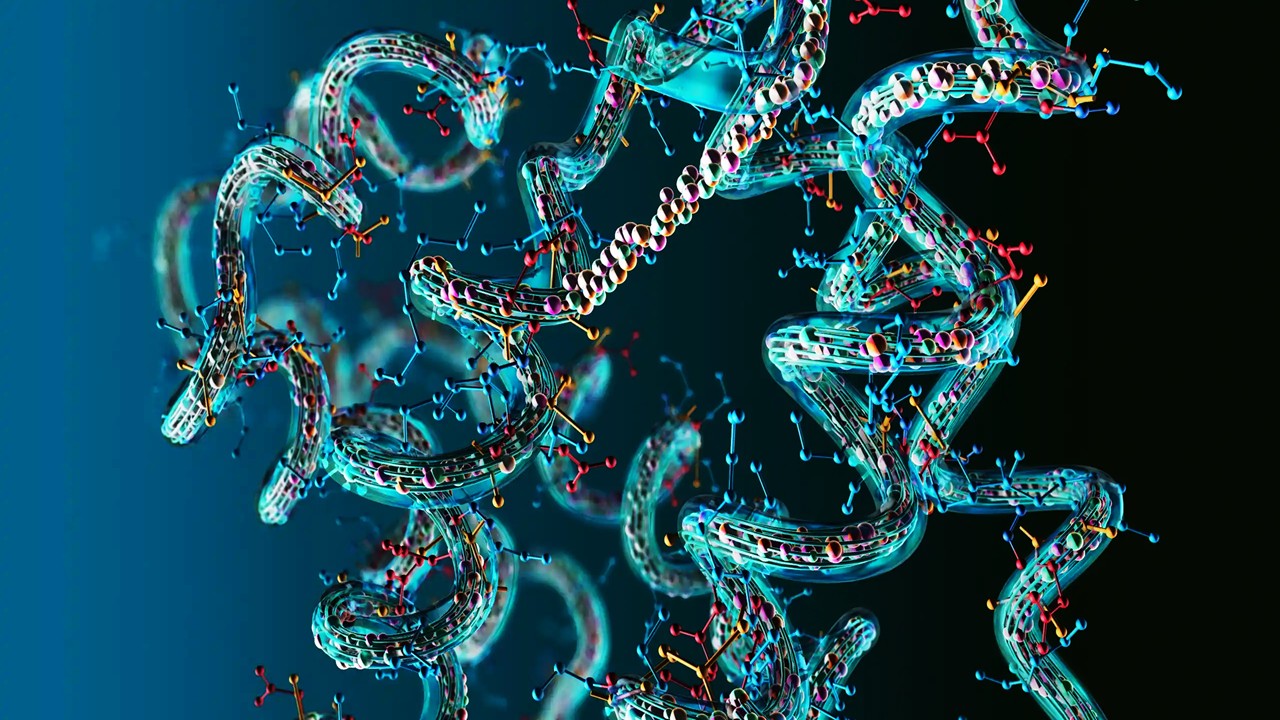
Semaphorin 3A (Sema3A), a neuronal guidance cue, emerges as a compelling candidate. Classically studied for axonal patterning, its functional expansion into vascular biology reframes the molecule as a bridge between nervous and circulatory systems. Elevated expression of Sema3A in the early weeks post-injury reveals a temporal alignment with nerve density peaks, suggesting it is not an incidental marker but a directional regulator. This observation directs inquiry toward the dualistic role of semaphorins as repellents and attractants, capable of sculpting not just nerve projections but endothelial tube formation. By mapping its co-localization with sensory nerve terminals, investigators link Sema3A release with downstream vascular clustering at the mineralizing front.
Thus, osteogenesis must be interpreted as an orchestrated event rather than a linear cascade. Sensory innervation establishes a biochemical niche, vascular structures follow, and mineral deposition consolidates this scaffold into functional tissue. This neurovascular interplay, anchored by Sema3A, introduces a redefinition of bone healing: bone is not simply rebuilt, it is rewired and revascularized in sequence. With this framework, the field shifts from describing osteogenesis as cell-centric to viewing it as network-centric, with nerves and vessels as its most vital infrastructural units.
Experimental Strategies for Visualizing Sequential Osteogenesis
The Achilles tenotomy rat model provides an accessible platform to study bone regeneration without implanting exogenous scaffolds. This model ensures that osteogenesis unfolds in situ, allowing endogenous regulators such as Sema3A to be traced in their natural kinetics. The rigor of experimental design rests on its multifaceted readouts, spanning structural imaging, histological staining, immunofluorescent mapping, and molecular quantification. These complementary methods converge to render a three-dimensional and temporal depiction of neurovascular dynamics within bone repair zones. Micro-computed tomography reveals density and volume changes, while immunofluorescence captures the molecular signatures that conventional imaging cannot resolve.
Micro-CT scans of the calcaneus and tibial insertion show mineral density increases over weeks, though the earliest time point lacks sufficient calcification for quantification. These findings suggest that bone mineral accrual is a lagging indicator compared to neural infiltration. Hematoxylin-eosin and Alizarin Red staining further confirm the emergence of trabecular structures by the sixth week, aligning histological detail with imaging-based quantification. This underscores the necessity of layering methodologies to avoid overlooking early neural or vascular events invisible to imaging thresholds. Only by cross-validating multiple modalities can temporal precedence between nerves, vessels, and bone be confidently established.
Immunofluorescence brings this precedence into sharp relief. CGRP-positive sensory nerves peak in density at the third week and then taper, while CD31- and endomucin-positive vessels intensify progressively toward the ninth week. This sequential shift is more than an observational curiosity—it signifies a regulated relay where neural infiltration hands off to vascular sprouting, which then enables osteoblast recruitment. Co-staining confirms colocalization between nerves and vessels, revealing intimate neurovascular partnerships rather than parallel growth trajectories. Such structural interdependencies make evident the necessity of nerve-derived molecules like Sema3A for establishing pro-angiogenic microenvironments.
Finally, qRT-PCR bridges structure with function by measuring gene expression signatures of sensory nerves, vessels, and osteogenic cells. Peaks of Cgrp and Sp in early weeks validate histological observations, while later surges in Cd31, Emcn, and osteogenic markers demonstrate vascular and mineral consolidation. Overlaying these with elevated Sema3a transcripts in dorsal root ganglia and injury sites closes the loop, linking neural infiltration to molecular guidance cues that orchestrate subsequent vascular and osteogenic events. Together, these strategies reconstruct osteogenesis as a sequentially tuned program.
Molecular Cartography of Sema3A and Sensory Nerve Influence
Semaphorin 3A holds center stage among neuronal guidance cues in this context. Unlike netrins or semaphorin 3E, whose expression remains relatively stable, Sema3A exhibits striking temporal elevation precisely when sensory nerves dominate the injury microenvironment. This temporal specificity suggests that Sema3A is not a passive correlate but an active effector, secreted by nerve terminals to condition their milieu. Immunofluorescent overlays confirm its co-localization with CGRP-positive fibers, further grounding the hypothesis that sensory nerves are the principal producers of Sema3A at osteogenic fronts.
The biological actions of Sema3A are paradoxical: it can repel axons in development yet attract endothelial sprouting under certain conditions. Within bone repair, this duality manifests as spatial steering—guiding nerves to establish gradients and directing endothelial progenitors to populate nerve-rich zones. Mechanistically, Sema3A interacts with neuropilin-1, competing with vascular endothelial growth factor to fine-tune angiogenic responses. In trauma-induced osteogenesis, this fine-tuning avoids chaotic vessel growth, ensuring that type H vessels form in close proximity to sensory nerves. These type H vessels, marked by dual CD31 and endomucin expression, are recognized as potent osteogenic enablers, linking Sema3A activity directly to mineralization outcomes.
Molecular assays strengthen this mechanistic link. Elevated Sema3a transcripts in dorsal root ganglia suggest central contributions to peripheral release, while localized protein expression within trauma sites affirms its bioactive distribution. Co-expression ratios of Sema3A and CGRP approach unity across experimental stages, indicating that nearly all sensory nerve terminals engaged in early infiltration are also Sema3A producers. This close correspondence underscores the molecule’s identity as a neurovascular mediator rather than an incidental marker. Such evidence situates Sema3A as both a spatial and temporal conductor of neurovascular assembly in bone repair.
By positioning sensory nerves as both structural infiltrators and molecular signalers, Sema3A reframes the neurovascular niche as an instructive rather than supportive environment. It challenges the notion that angiogenesis and mineralization are self-organizing, instead proposing a nerve-dependent cascade. These insights create new interpretive maps of bone healing, where Sema3A gradients chart vascular territories and sensory fibers establish the blueprint for osteogenesis. The emerging picture is one of bone repair as guided tissue assembly rather than spontaneous regeneration.
From Sequential Dynamics to Osteogenic Consolidation
The temporal unfolding of osteogenesis begins with neural infiltration, transitions to vascular sprouting, and culminates in mineral consolidation. Each phase is not isolated but sequentially dependent, with preceding stages priming the microenvironment for subsequent events. Sensory nerves dominate the earliest phase, peaking in density before vessel sprouting intensifies. Their withdrawal over time does not mark irrelevance but rather the completion of their instructive role. What remains is a scaffold enriched with molecular cues like Sema3A that continues to direct vascular and osteogenic activity even as nerve density declines.
Angiogenesis, particularly the formation of type H vessels, represents the second phase. These vessels differ from general capillaries by their close coupling with osteoblastic activity and higher capacity to promote bone mass accrual. Their co-localization with early neural tracts highlights the precision of spatial signaling. Sema3A again mediates this by enabling endothelial responsiveness to osteogenic needs while tempering excessive sprouting. The consequence is vascular growth that aligns with mineralization fronts, avoiding disorganized networks that could hinder structural consolidation.
Mineralization defines the final phase, consolidating the neurovascular groundwork into calcified architecture. Micro-CT and histological markers confirm that mineral density continues to rise even as nerve density falls, illustrating the handoff between early neural instruction and long-term osteogenic execution. At this point, osteoblast differentiation markers peak, supported by the vascular supply established in the preceding phase. The interdependence between nerves, vessels, and bone is therefore sequential but also cyclical, with feedback loops maintaining equilibrium. Bone is not a static endpoint but a dynamically remodeled tissue whose formation replays these neurovascular sequences at every repair cycle.
The recognition of sequential neurovascular dynamics broadens the therapeutic lens for skeletal disorders. Interventions that ignore early neural infiltration risk missing critical windows where sensory nerves set the trajectory for healing. Conversely, targeting Sema3A or related pathways may allow clinicians to extend or enhance this instructive phase, accelerating angiogenesis and mineralization in trauma or degenerative conditions. By framing bone healing as an orchestrated sequence rather than a simple accumulation of cells, researchers open new avenues for modulation that align with natural regenerative programming.
Implications for Regenerative Medicine and Therapeutic Design
The identification of Sema3A as a bioactive regulator of sequential osteogenesis carries translational significance. Bone repair strategies, whether grafts, scaffolds, or biologics, often focus on osteoblast recruitment and angiogenic induction as separate interventions. Yet, this study reveals that without the prior neural phase and its molecular cues, these downstream processes lack organization. Incorporating neurotrophic signaling, particularly Sema3A pathways, could refine current approaches by embedding neurovascular orchestration into biomaterial design. Scaffold systems infused with Sema3A mimetics may precondition vascular alignment before mineralization ensues.
Beyond biomaterials, pharmacological interventions targeting Sema3A signaling open therapeutic frontiers. Agents that modulate neuropilin-1 binding or Sema3A release from dorsal root ganglia could be harnessed to accelerate or amplify bone repair. Such approaches differ from conventional osteoanabolic drugs by acting not directly on osteoblasts but on the neurovascular niche that governs their function. This represents a paradigm shift where nerve-derived molecules are seen as primary therapeutic targets rather than peripheral modulators. The implications extend to fractures, nonunions, and even metabolic bone diseases where disrupted neurovascular coordination undermines repair.
The findings also resonate with broader fields of regenerative biology. Neurovascular coupling is fundamental not only in bone but in skin, muscle, and even cardiac repair. By delineating the sequential order in bone healing, this work provides a transferable framework for other tissues where sensory nerves and vessels co-inhabit regenerative landscapes. It challenges the field to consider how neuronal guidance cues could be therapeutically mobilized in diverse contexts. The universality of semaphorin biology makes such cross-tissue applications plausible, broadening the reach of these discoveries beyond orthopedics.
In summary, the sequential formation of nerves and type H vessels, orchestrated by Sema3A, elevates sensory innervation from a supportive role to a commanding one in osteogenesis. The recognition of this hierarchy redefines how we understand skeletal regeneration and how we might design interventions to accelerate or enhance it. By targeting the earliest cues, medicine can potentially harness the full regenerative capacity of bone, integrating nerves, vessels, and minerals into a synchronized architecture of repair.
Study DOI: https://doi.org/10.3389/fbioe.2023.1138601
Engr. Dex Marco Tiu Guibelondo, B.Sc. Pharm, R.Ph., B.Sc. CpE
Editor-in-Chief, PharmaFEATURES

Subscribe
to get our
LATEST NEWS
Related Posts
Regenerative Medicine & Biomaterials
Programmable Nitric Grafts: Steady Nitric Oxide Orchestrates Small-Vessel Healing
Embedding nitrate-driven nitric-oxide release within PCL grafts programmatically guides vascular regeneration across the lumen, media, and progenitor niche.

Regenerative Medicine & Biomaterials
Bioactive Networks: Photocrosslinkable Gelatin Hydrogels and Cytokine Modulation in Human Immunity
GelMA hydrogels demonstrate that the architecture of matter itself can modulate the architecture of inflammation.

Read More Articles


Medicinal Chemistry & Pharmacology
April 14, 2026
Igor Nasonkin and Phythera Therapeutics: Moving Oncology Beyond Single Targets into Engineered Polypharmacologic Systems
Igor Nasonkin’s systems-driven approach at Phythera Therapeutics reframes oncology drug development from single-target inhibition to AI-enabled polypharmacologic network modulation using nature-derived molecular architectures.


Artificial Intelligence and Data Analytics
April 10, 2026
Inside Johnson & Johnson’s External Innovation Engine: Devin Swanson on Translating Integrated Discovery into Strategic Value
Devin Swanson’s leadership at Johnson & Johnson Innovative Medicines redefines external innovation as a tightly governed, AI-enabled translational system integrating multi-modal drug discovery, biomarker strategy, and capital-efficient execution.

Immunology & Oncology
April 9, 2026
From DMPK to Distributed Execution: Mehran F. Moghaddam’s Systems Strategy at OROX BioSciences, Inc.
A systems-level examination of how Mehran F. Moghaddam operationalizes DMPK, externalized R&D, and lipid-mediated therapeutics into a predictive, high-velocity biotech development architecture.